 Explore
 Validate
 Learn
Learn Western blot
Antibody data
- Antibody Data
- Antigen structure
- References [0]
- Comments [0]
- Validations
- Western blot [1]
- Immunocytochemistry [1]
- Flow cytometry [1]
Submit
Validation data
Reference
Comment
Report error
- Product number
- PA5-119942 - Provider product page

- Provider
- Invitrogen Antibodies
- Product name
- LRRTM1 Polyclonal Antibody
- Antibody type
- Polyclonal
- Antigen
- Synthetic peptide
- Reactivity
- Human, Mouse, Rat
- Host
- Rabbit
- Isotype
- IgG
- Vial size
- 100 µL
- Concentration
- 1 mg/mL
- Storage
- Store at 4°C short term. For long term storage, store at -20°C, avoiding freeze/thaw cycles.
No comments: Submit comment
Supportive validation
- Submitted by
- Invitrogen Antibodies (provider)
- Main image
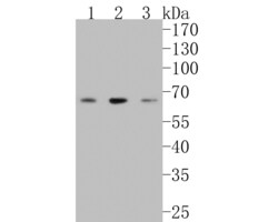
- Experimental details
- Western blot analysis of LRRTM1 on different lysates. Proteins were transferred to a PVDF membrane and blocked with 5% NFDM/TBST for 1 hour at room temperature. LRRTM1 Polyclonal Antibody (Product # PA5-119942) at 1:500 was used in 5% NFDM/TBST at room temperature for 2 hours. Goat Anti-Rabbit IgG - HRP Secondary Antibody at 1:200,000 dilution was used for 1 hour at room temperature. Positive control: Lane 1: SH-SY5Y cell lysate. Lane 2: N2A cell lysate. Lane 1: Rat brain tissue lysate.

Supportive validation
- Submitted by
- Invitrogen Antibodies (provider)
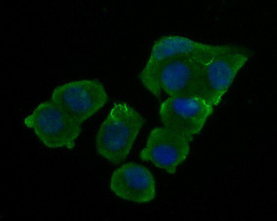
- Main image

- Experimental details
- Immunocytochemistry-Immunofluorescence analysis of of LRRTM1 in N2A cells (green). Formalin fixed cells were permeabilized with 0.1% Triton X-100 in TBS for 10 minutes at room temperature and blocked with 10% negative goat serum for 15 minutes at room temperature. Cells were probed with LRRTM1 Polyclonal Antibody (Product # PA5-119942) at a dilution of 1:200 for 1 hour at room temperature, washed with PBS. Alexa Fluor 488 Goat anti-Rabbit IgG was used as the secondary antibody at 1:1,000 dilution. The nuclear counter stain is DAPI (blue).
Supportive validation
- Submitted by
- Invitrogen Antibodies (provider)
- Main image

- Experimental details
- Flow Cytometry analysis of LRRTM1 in SH-SY5Y cells using LRRTM1 Polyclonal Antibody (Product # PA5-119942) at 1 µg/mL (red) compared with Rabbit IgG, monoclonal - Isotype Control (green). After incubation of the primary antibody at 4°C for 1 hour, the cells were stained with a Alexa Fluor 488 conjugate-Goat anti-Rabbit IgG Secondary antibody at 1:1,000 dilution for 30 minutes at 4°C (dark incubation). Unlabelled sample was used as a control (cells without incubation with primary antibody; black).
