Explore
 Validate
 Learn
LearnMAB7774R-100
antibody from R&D Systems
Targeting: IL1F10
FKSG75, IL-1F10, IL-1HY2, IL1-theta, MGC11983, MGC119832, MGC119833
 Western blot
Antibody data
- Antibody Data
- Antigen structure
- References [0]
- Comments [0]
- Validations
- Western blot [1]
- ELISA [1]
- Flow cytometry [1]
Submit
Validation data
Reference
Comment
Report error
- Product number
- MAB7774R-100 - Provider product page

- Provider
- R&D Systems
- Product name
- Mouse IL-38/IL-1F10 Antibody
- Antibody type
- Monoclonal
- Description
- Protein A or G purified from cell culture supernatant. Detects mouse IL-38/IL-1F10 in direct ELISAs and Western blots.
- Reactivity
- Mouse
- Host
- Rat
- Conjugate
- Unconjugated
- Antigen sequence
Q8R459- Isotype
- IgG
- Antibody clone number
- 798036R
- Vial size
- 100 ug
- Storage
- Use a manual defrost freezer and avoid repeated freeze-thaw cycles. 12 months from date of receipt, -20 to -70 °C as supplied. 1 month, 2 to 8 °C under sterile conditions after reconstitution. 6 months, -20 to -70 °C under sterile conditions after reconstitution.
No comments: Submit comment
Supportive validation
- Submitted by
- R&D Systems (provider)
- Main image
- Experimental details
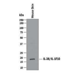
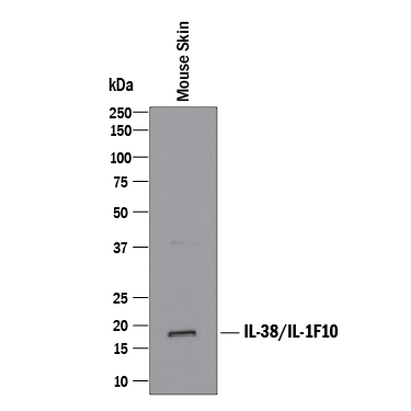
- Detection of Mouse IL-38/IL-1F10 by Western Blot. Western blot shows lysates of mouse skin tissue. PVDF membrane was probed with 2 µg/mL of Recombinant Rat Anti-Mouse IL-38/IL-1F10 Monoclonal Antibody (Catalog # MAB7774R) followed by HRP-conjugated Anti-Rat IgG Secondary Antibody (Catalog # HAF005). A specific band was detected for IL-38/IL-1F10 at approximately 18 kDa (as indicated). This experiment was conducted under reducing conditions and using Immunoblot Buffer Group 1.
Supportive validation
- Submitted by
- R&D Systems (provider)
- Main image
- Experimental details
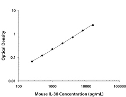
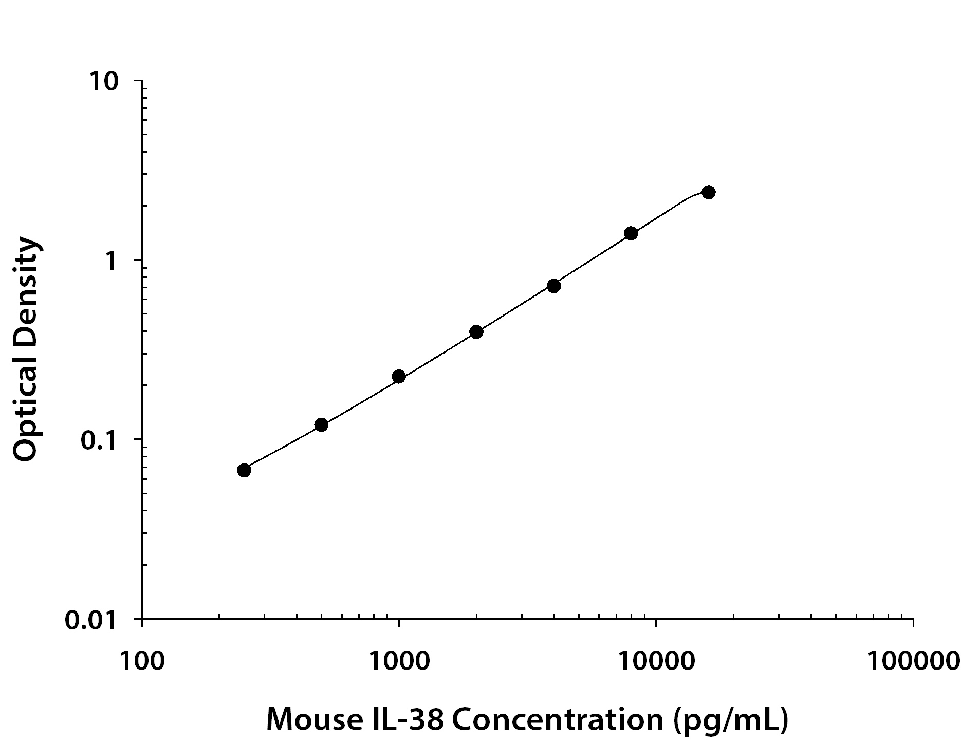
- Mouse IL-38/IL-1F10 ELISA Standard Curve. Recombinant Mouse IL-38/IL-1F10 protein was serially diluted 2-fold and captured by Rat Anti-Mouse IL-38/IL-1F10 Monoclonal Antibody (Catalog # MAB77742) coated on a Clear Polystyrene Microplate (Catalog # DY990). Rat Anti-Mouse IL-38/IL-1F10 Monoclonal Antibody (Catalog # MAB7774R) was biotinylated and incubated with the protein captured on the plate. Detection of the standard curve was achieved by incubating Streptavidin-HRP (Catalog # DY998) followed by Substrate Solution (Catalog # DY999) and stopping the enzymatic reaction with Stop Solution (Catalog # DY994).
Supportive validation
- Submitted by
- R&D Systems (provider)
- Main image
- Experimental details
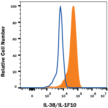
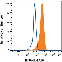
- Detection of IL-38/IL-1F10 in RAW 264.7 Mouse Cell Line by Flow Cytometry. RAW 264.7 mouse monocyte/macrophage cell line was stained with Recombinant Rat Anti-Mouse IL-38/IL-1F10 Monoclonal Antibody (Catalog # MAB7774R, filled histogram) or isotype control antibody (Catalog # MAB006, open histogram), followed by Fluorescein-conjugated Anti-Rat IgG Secondary Antibody (Catalog # F0104B). To facilitate intracellular staining, cells were fixed with Flow Cytometry Fixation Buffer (Catalog # FC004) and permeabilized with Flow Cytometry Permeabilization/Wash Buffer I (Catalog # FC005). View our protocol for Staining Intracellular Molecules.