Explore
 Validate
 Learn
LearnPA5-27077
antibody from Invitrogen Antibodies
Targeting: ACKR3
CMKOR1, CXCR7, GPR159, RDC1
 Western blot Immunocytochemistry
 Immunoprecipitation Immunohistochemistry Flow cytometry Other assay
Antibody data
- Antibody Data
- Antigen structure
- References [2]
- Comments [0]
- Validations
- Western blot [5]
- Immunocytochemistry [1]
- Immunohistochemistry [3]
- Other assay [1]
Submit
Validation data
Reference
Comment
Report error
- Product number
- PA5-27077 - Provider product page

- Provider
- Invitrogen Antibodies
- Product name
- CXCR7 Polyclonal Antibody
- Antibody type
- Polyclonal
- Antigen
- Synthetic peptide
- Reactivity
- Human, Mouse
- Host
- Rabbit
- Isotype
- IgG
- Vial size
- 100 µL
- Concentration
- 0.14 mg/mL
- Storage
- Store at 4°C short term. For long term storage, store at -20°C, avoiding freeze/thaw cycles.
Submitted references Selective Elimination of Senescent Fibroblasts by Targeting the Cell Surface Protein ACKR3.
Exploration of biomarkers from a pilot weight management study for men undergoing radical prostatectomy.
Takaya K, Asou T, Kishi K
International journal of molecular sciences 2022 Jun 10;23(12)
International journal of molecular sciences 2022 Jun 10;23(12)
Exploration of biomarkers from a pilot weight management study for men undergoing radical prostatectomy.
Dimachkie MD, Bechtel MD, Robertson HL, Michel C, Lee EK, Sullivan DK, Chalise P, Thrasher JB, Parker WP, Godwin AK, Pathak HB, DiGiovanni J, Shivappa N, Hébert JR, Hamilton-Reeves JM
Urologic oncology 2021 Aug;39(8):495.e7-495.e15
Urologic oncology 2021 Aug;39(8):495.e7-495.e15
No comments: Submit comment
Supportive validation
- Submitted by
- Invitrogen Antibodies (provider)
- Main image
- Experimental details
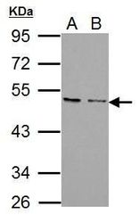
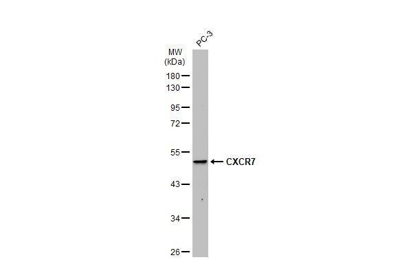
- Western blot analysis of CXCR7 using 30 µg of A) PC-3 and B) U87-MG lysate. Samples were loaded onto a 10% SDS-PAGE gel and probed with a CXCR7 polyclonal antibody (Product # PA5-27077) at a dilution of 1:1000.
- Submitted by
- Invitrogen Antibodies (provider)
- Main image
- Experimental details
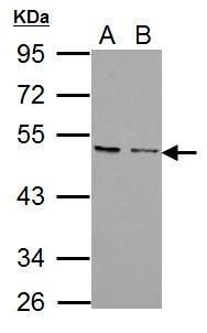
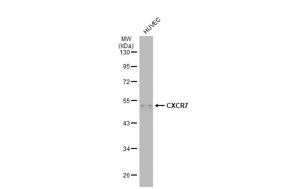
- Western Blot using CXCR7 Polyclonal Antibody (Product # PA5-27077). Whole cell extract (30 µg) was separated by 10% SDS-PAGE, and the membrane was blotted with CXCR7 Polyclonal Antibody (Product # PA5-27077) diluted at 1:1,000. The HRP-conjugated anti-rabbit IgG antibody was used to detect the primary antibody, and the signal was developed with Trident ECL plus-Enhanced.
- Submitted by
- Invitrogen Antibodies (provider)
- Main image
- Experimental details
- Western Blot using CXCR7 Polyclonal Antibody (Product # PA5-27077). Whole cell extract (30 µg) was separated by 10% SDS-PAGE, and the membrane was blotted with CXCR7 Polyclonal Antibody (Product # PA5-27077) diluted at 1:500. The HRP-conjugated anti-rabbit IgG antibody was used to detect the primary antibody.
- Submitted by
- Invitrogen Antibodies (provider)
- Main image
- Experimental details
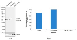
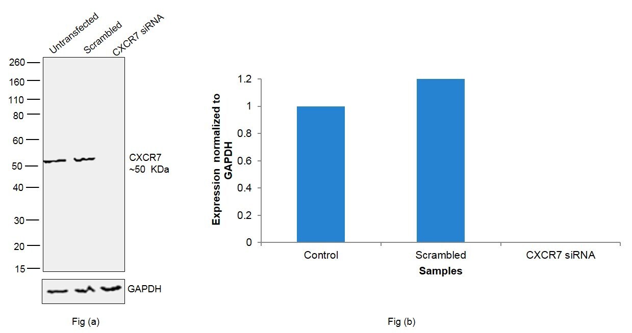
- Knockdown of CXCR7 was achieved by transfecting LNCaP cells with CXCR7specific siRNAs (Silencer® select Product # s94, s95). Western blot analysis (Fig. a) was performed using whole cell extracts from the CXCR7 knockdown cells (lane 3), non-specific scrambled siRNA transfected cells (lane 2) and untransfected cells (lane 1). The blot was probed with CXCR7 Polyclonal Antibody (Product # PA5-27077, 1:1000 dilution) and Goat anti-Rabbit IgG (H+L), Superclonal™ Recombinant Secondary Antibody, HRP (Product # A27036, 1:4000 dilution). Densitometric analysis of this western blot is shown in histogram (Fig. b). Decrease in signal upon siRNA mediated knock down confirms that antibody is specific to CXCR7..
- Submitted by
- Invitrogen Antibodies (provider)
- Main image
- Experimental details
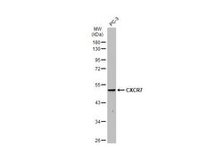
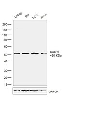
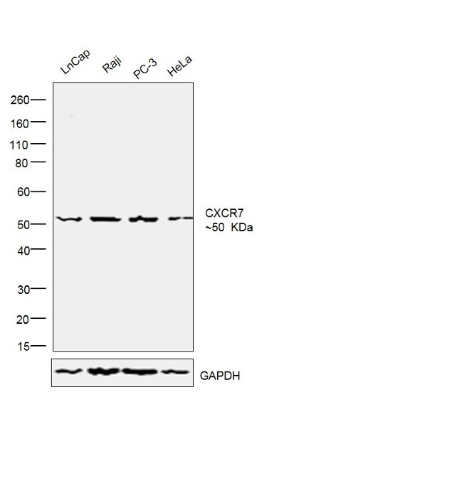
- Western blot was performed using Anti-CXCR7 polyclonal Antibody (Product # PA5-27077) and ~50kDa band corresponding to CXCR7 was observed across the cell lines tested. Whole cell extracts (30ug lysate) (1% SDS) of LNCaP (Lane 1), Raji (Lane 2), PC-3 (Lane 3) and HeLa (Lane 4) were electrophoresed using Novex® NuPAGE® 4-12 % Bis-Tris gel (Product # NP0322BOX). Resolved proteins were then transferred onto a nitrocellulose membrane (Product # IB23001) by iBlot® 2 Dry Blotting System (Product # IB21001). The blot was probed with the primary antibody (1:1000 dilution) and detected by chemiluminescence with Goat anti-Rabbit IgG (H+L) Superclonal™ Recombinant Secondary Antibody, HRP (Product # A27036, 1:4000 dilution) using the iBright FL 1000 (Product # A32752). Chemiluminescent detection was performed using Novex® ECL Chemiluminescent Substrate Reagent Kit (Product # WP20005)..
Supportive validation
- Submitted by
- Invitrogen Antibodies (provider)
- Main image
- Experimental details
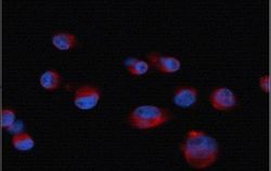
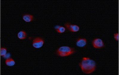
- Immunofluorescent analysis of CXCR7 in paraformaldehyde-fixed human pancreatic cancer cell line cells using a CXCR7 polyclonal antibody (Product # PA5-27077) at a 1:50 dilution. Blue: DAPI.
Supportive validation
- Submitted by
- Invitrogen Antibodies (provider)
- Main image
- Experimental details
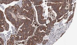
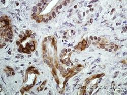
- Immunohistochemical analysis of paraffin-embedded human gastric cancer, using CXCR7 (Product # PA5-27077) antibody at 1:100 dilution. Antigen Retrieval: EDTA based buffer, pH 8.0, 15 min.
- Submitted by
- Invitrogen Antibodies (provider)
- Main image
- Experimental details
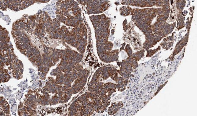
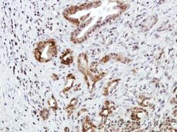
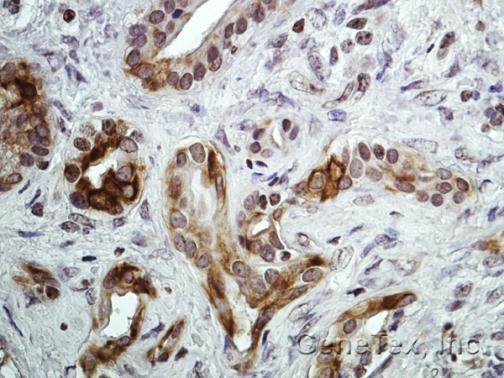
- Immunohistochemical analysis of paraffin-embedded Human pancreatic tumor, using CXCR7 (Product # PA5-27077) antibody at 1:100 dilution. Antigen Retrieval: EDTA based buffer, pH 8.0, 15 min.
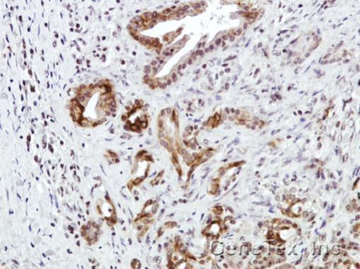
- Submitted by
- Invitrogen Antibodies (provider)
- Main image
- Experimental details
- Immunohistochemical analysis of paraffin-embedded Human pancreatic tumor, using CXCR7 (Product # PA5-27077) antibody at 1:100 dilution. Antigen Retrieval: EDTA based buffer, pH 8.0, 15 min.
Supportive validation
- Submitted by
- Invitrogen Antibodies (provider)
- Main image
- Experimental details
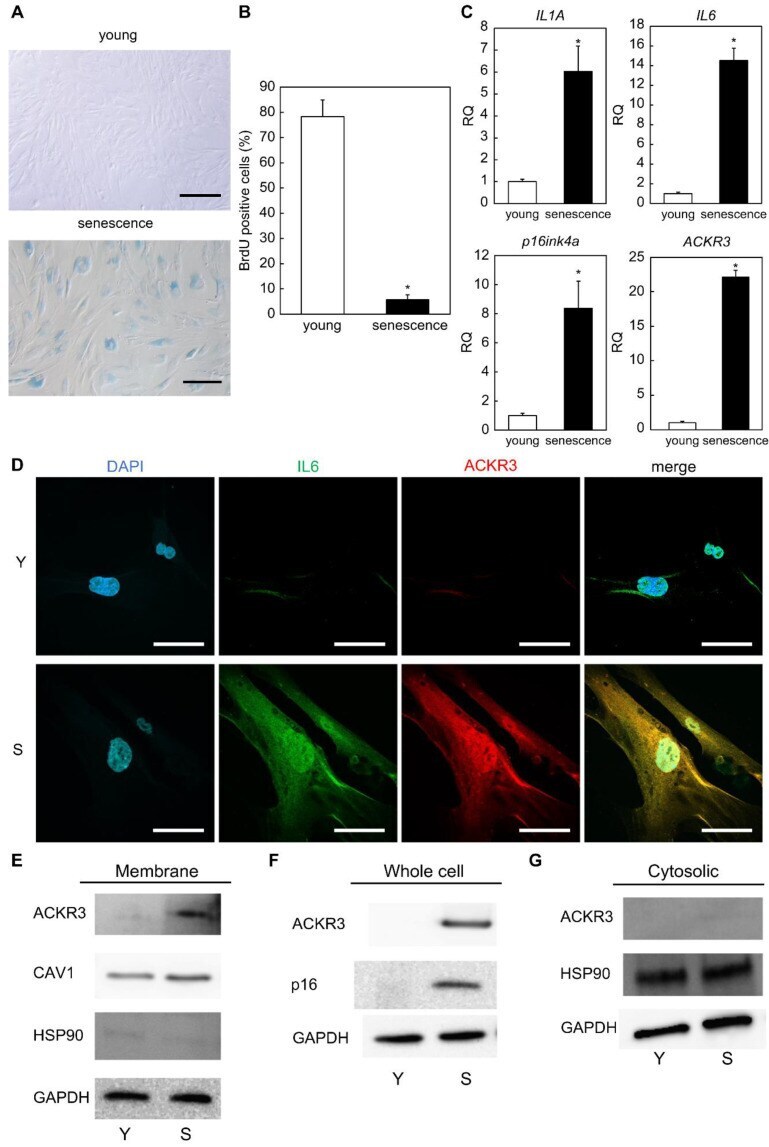
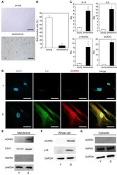
- Identification of ACKR3 as a novel senescent cell surface marker protein. ( A ) SA-beta-gal staining of proliferating and senescent cells. Bar = 50 um. ( B ) BrdU absorption by proliferating and senescent cells. ( C ) Real-time quantitative polymerase chain reaction (RT-qPCR) analysis of the expression of senescence-related genes using cell extracts. GAPDH was used as the housekeeping gene. ( D ) Immunostaining of IL6 and ACKR3 in young (proliferating) and senescent cells. Bar = 20 um. Y: young cells. S: senescent cells. ( E ) Western blot analysis of cell extract proteins. ( F ) Western blot analysis of whole-cell lysate proteins. ( G ) Western blot analysis of cytosolic proteins. GAPDH was used as the housekeeping protein. *; p < 0.05. RQ; relative quantification. All experiments were repeated in triplicate.