 Explore
 Validate
 Learn
Learn Immunohistochemistry
Antibody data
- Antibody Data
- Antigen structure
- References [4]
- Comments [0]
- Validations
- Immunohistochemistry [2]
- Flow cytometry [1]
Submit
Validation data
Reference
Comment
Report error
- Product number
- MAB4227 - Provider product page

- Provider
- R&D Systems
- Product name
- Human CXCR7/RDC-1 Antibody
- Antibody type
- Monoclonal
- Description
- Protein A or G purified from hybridoma culture supernatant. Detects human CXCR7/RDC-1 in direct ELISAs. In flow cytometry, reacts specifically with five distinct human CXCR7 transfectants, but does not react with their respective parental lines or mouse CXCR7 transfectants. In flow cytometry, also reacts with monocytes expressing CXCR7, but does not react with MCF-7 cells which have been reported to have surface-expressing CXCR7 using clone 11G8. Due to the conflicting reports published, use of monoclonal MAB4227 may result in an underestimation of CXCR7 expression on certain cell types.
- Reactivity
- Human
- Host
- Mouse
- Conjugate
- Unconjugated
- Antigen sequence
AAA62370- Isotype
- IgG
- Antibody clone number
- 358426
- Vial size
- 100 ug
- Concentration
- LYOPH
- Storage
- Use a manual defrost freezer and avoid repeated freeze-thaw cycles. 12 months from date of receipt, -20 to -70 °C as supplied. 1 month, 2 to 8 °C under sterile conditions after reconstitution. 6 months, -20 to -70 °C under sterile conditions after reconstitution.
Submitted references Ligand-specific conformational transitions and intracellular transport are required for atypical chemokine receptor 3-mediated chemokine scavenging.
Isolation and characterization of human trophoblast side-population (SP) cells in primary villous cytotrophoblasts and HTR-8/SVneo cell line.
Analysis of HLDA9 mAbs on plasmacytoid dendritic cells.
CXCR7 protein is not expressed on human or mouse leukocytes.
Montpas N, St-Onge G, Nama N, Rhainds D, Benredjem B, Girard M, Hickson G, Pons V, Heveker N
The Journal of biological chemistry 2018 Jan 19;293(3):893-905
The Journal of biological chemistry 2018 Jan 19;293(3):893-905
Isolation and characterization of human trophoblast side-population (SP) cells in primary villous cytotrophoblasts and HTR-8/SVneo cell line.
Takao T, Asanoma K, Kato K, Fukushima K, Tsunematsu R, Hirakawa T, Matsumura S, Seki H, Takeda S, Wake N
PloS one 2011;6(7):e21990
PloS one 2011;6(7):e21990
Analysis of HLDA9 mAbs on plasmacytoid dendritic cells.
Cabezón R, Sintes J, Llinàs L, Benitez-Ribas D
Immunology letters 2011 Jan 30;134(2):167-73
Immunology letters 2011 Jan 30;134(2):167-73
CXCR7 protein is not expressed on human or mouse leukocytes.
Berahovich RD, Zabel BA, Penfold ME, Lewén S, Wang Y, Miao Z, Gan L, Pereda J, Dias J, Slukvin II, McGrath KE, Jaen JC, Schall TJ
Journal of immunology (Baltimore, Md. : 1950) 2010 Nov 1;185(9):5130-9
Journal of immunology (Baltimore, Md. : 1950) 2010 Nov 1;185(9):5130-9
No comments: Submit comment
Supportive validation
- Submitted by
- R&D Systems (provider)
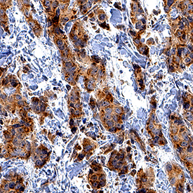
- Main image

- Experimental details
- CXCR7/RDC-1 in Human Breast Cancer Tissue. CXCR7/RDC-1 was detected in immersion fixed paraffin-embedded sections of human breast cancer tissue using Mouse Anti-Human CXCR7/RDC-1 Monoclonal Antibody (Catalog # MAB4227) at 0.5 µg/mL for 1 hour at room temperature followed by incubation with the Anti-Mouse IgG VisUCyte™ HRP Polymer Antibody (Catalog # VC001). Tissue was stained using DAB (brown) and counterstained with hematoxylin (blue). Specific staining was localized to cytoplasm and plasma membrane. View our protocol for IHC Staining with VisUCyte HRP Polymer Detection Reagents.
- Submitted by
- R&D Systems (provider)
- Main image
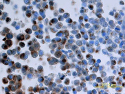
- Experimental details
- CXCR7/RDC-1 in Human Breast Cancer Tissue. CXCR7/RDC-1 was detected in perfusion fixed paraffin-embedded sections of nude mice injected with human breast cancer cells using Mouse Anti-Human CXCR7/RDC-1 Monoclonal Antibody (Catalog # MAB4227) at 25 µg/mL overnight at 4 °C. Tissue was stained using the Anti-Mouse HRP-DAB Cell & Tissue Staining Kit (brown; Catalog # CTS002) and counterstained with hematoxylin (blue). View our protocol for Chromogenic IHC Staining of Paraffin-embedded Tissue Sections.

Supportive validation
- Submitted by
- R&D Systems (provider)
- Main image
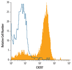
- Experimental details
- Detection of CXCR7/RDC-1 in Human peripheral blood Monocytes by Flow Cytometry. Human peripheral blood monocytes were stained with Mouse Anti-Human CXCR7/RDC-1 Monoclonal Antibody (Catalog # MAB4227, filled histogram) or isotype control antibody (Catalog # MAB003, open histogram), followed by Allophycocyanin-conjugated Anti-Mouse IgG F(ab')2 Secondary Antibody (Catalog # F0101B).
