 Explore
 Validate
 Learn
Learn Western blot
Antibody data
- Antibody Data
- Antigen structure
- References [2]
- Comments [0]
- Validations
- Western blot [3]
- Immunohistochemistry [1]
- Blocking/Neutralizing [1]
Submit
Validation data
Reference
Comment
Report error
- Product number
- AF1074 - Provider product page

- Provider
- R&D Systems
- Product name
- Human/Mouse JAM-B/VE-JAM Antibody
- Antibody type
- Polyclonal
- Description
- Antigen Affinity-purified. Detects human JAM-B/VE-JAM in direct ELISAs. Detects human and mouse JAM-B/VE-JAM in Western blots.
- Reactivity
- Human, Mouse
- Host
- Goat
- Conjugate
- Unconjugated
- Antigen sequence
P57087- Isotype
- IgG
- Vial size
- 100 ug
- Concentration
- LYOPH
- Storage
- Use a manual defrost freezer and avoid repeated freeze-thaw cycles. 12 months from date of receipt, -20 to -70 °C as supplied. 1 month, 2 to 8 °C under sterile conditions after reconstitution. 6 months, -20 to -70 °C under sterile conditions after reconstitution.
Submitted references Junctional adhesion molecule (JAM)-B supports lymphocyte rolling and adhesion through interaction with alpha4beta1 integrin.
Possible involvement of gap junctions in the barrier function of tight junctions of brain and lung endothelial cells.
Ludwig RJ, Hardt K, Hatting M, Bistrian R, Diehl S, Radeke HH, Podda M, Schön MP, Kaufmann R, Henschler R, Pfeilschifter JM, Santoso S, Boehncke WH
Immunology 2009 Oct;128(2):196-205
Immunology 2009 Oct;128(2):196-205
Possible involvement of gap junctions in the barrier function of tight junctions of brain and lung endothelial cells.
Nagasawa K, Chiba H, Fujita H, Kojima T, Saito T, Endo T, Sawada N
Journal of cellular physiology 2006 Jul;208(1):123-32
Journal of cellular physiology 2006 Jul;208(1):123-32
No comments: Submit comment
Supportive validation
- Submitted by
- R&D Systems (provider)
- Main image

- Experimental details
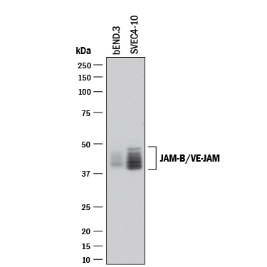
- Detection of Mouse JAM-B/VE-JAM by Western Blot. Western blot shows lysates of bEnd.3 mouse endothelioma cell line and SVEC4-10 mouse vascular endothelial cell line. PVDF membrane was probed with 2 µg/mL of Goat Anti-Human/Mouse JAM-B/VE-JAM Antigen Affinity-purified Polyclonal Antibody (Catalog # AF1074) followed by HRP-conjugated Anti-Goat IgG Secondary Antibody (Catalog # HAF017). Specific bands were detected for JAM-B/VE-JAM at approximately 40-48 kDa (as indicated). This experiment was conducted under reducing conditions and using Immunoblot Buffer Group 1.
- Submitted by
- R&D Systems (provider)
- Main image
- Experimental details
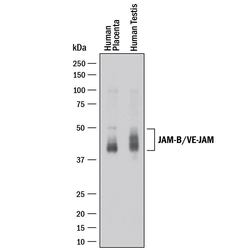
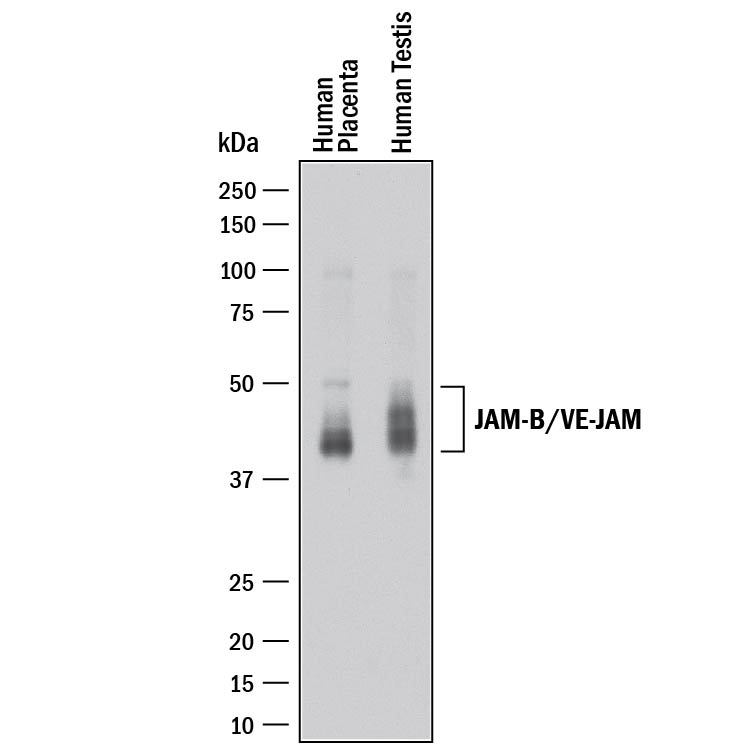
- Detection of Human JAM-B/VE-JAM by Western Blot. Western blot shows lysates of human placenta tissue and human testis tissue. PVDF membrane was probed with 1 µg/mL of Goat Anti-Human/Mouse JAM-B/VE-JAM Antigen Affinity-purified Polyclonal Antibody (Catalog # AF1074) followed by HRP-conjugated Anti-Goat IgG Secondary Antibody (Catalog # HAF017). Specific bands were detected for JAM-B/VE-JAM at approximately 43-50 kDa (as indicated). This experiment was conducted under reducing conditions and using Immunoblot Buffer Group 1.
- Submitted by
- R&D Systems (provider)
- Main image

- Experimental details
- Detection of Human JAM-B/VE-JAM by Simple WesternTM. Simple Western lane view shows lysates of human placenta tissue and human testis tissue, loaded at 0.2 mg/mL. A specific band was detected for JAM-B/VE-JAM at approximately 58-61 kDa (as indicated) using 20 µg/mL of Goat Anti-Human JAM-B/VE-JAM Antigen Affinity-purified Polyclonal Antibody (Catalog # AF1074) followed by 1:50 dilution of HRP-conjugated Anti-Goat IgG Secondary Antibody (Catalog # HAF109). This experiment was conducted under reducing conditions and using the 12-230 kDa separation system.

Supportive validation
- Submitted by
- R&D Systems (provider)
- Main image

- Experimental details
- JAM-B/VE-JAM in Human Tonsil. JAM-B/VE-JAM was detected in immersion fixed paraffin-embedded sections of human tonsil using Goat Anti-Human/Mouse JAM-B/VE-JAM Antigen Affinity-purified Polyclonal Antibody (Catalog # AF1074) at 3 µg/mL for 1 hour at room temperature followed by incubation with the Anti-Goat IgG VisUCyte™ HRP Polymer Antibody (Catalog # VC004). Tissue was stained using DAB (brown) and counterstained with hematoxylin (blue). Specific staining was localized to plasma membrane in endothelial cells. View our protocol for IHC Staining with VisUCyte HRP Polymer Detection Reagents.

Supportive validation
- Submitted by
- R&D Systems (provider)
- Main image

- Experimental details
- Cell Adhesion Mediated by JAM-B/VE-JAM and Neutralization by Human JAM-B/VE-JAM Antibody. Recombinant Human JAM-B/VE-JAM Fc Chimera (Catalog # 1074-VJ), immobilized onto a microplate previously coated with Goat Anti-Human IgG Fc (Catalog # G-102-C), supports the adhesion of the J45.01 human acute lymphoblastic leukemia T lymphocyte cell line in a dose-dependent manner (orange line), as measured by endogenous cellular lysosomal acid phosphatase activity. Adhesion elicited by Recombinant Human JAM-B/VE-JAM Fc Chimera (0.2 µg/mL) is neutralized (green line) by increasing concentrations of Goat Anti-Human/Mouse JAM-B/VE-JAM Antigen Affinity-purified Polyclonal Antibody (Catalog # AF1074). The ND50 is typically 0.2-0.8 µg/mL.
