Explore
 Validate
 Learn
Learn Western blot
Antibody data
- Antibody Data
- Antigen structure
- References [0]
- Comments [0]
- Validations
- Western blot [2]
- Immunohistochemistry [1]
Submit
Validation data
Reference
Comment
Report error
- Product number
- PA5-47638 - Provider product page

- Provider
- Invitrogen Antibodies
- Product name
- TENM2 Polyclonal Antibody
- Antibody type
- Polyclonal
- Antigen
- Recombinant full-length protein
- Description
- In direct ELISAs, approximately 15% cross-reactivity with recombinant human (rh) Teneurin-3 is observed, and less than 4% cross-reactivity with rhTeneurin-1 is observed.
- Concentration
- 0.2 mg/mL
No comments: Submit comment
Supportive validation
- Submitted by
- Invitrogen Antibodies (provider)
- Main image

- Experimental details
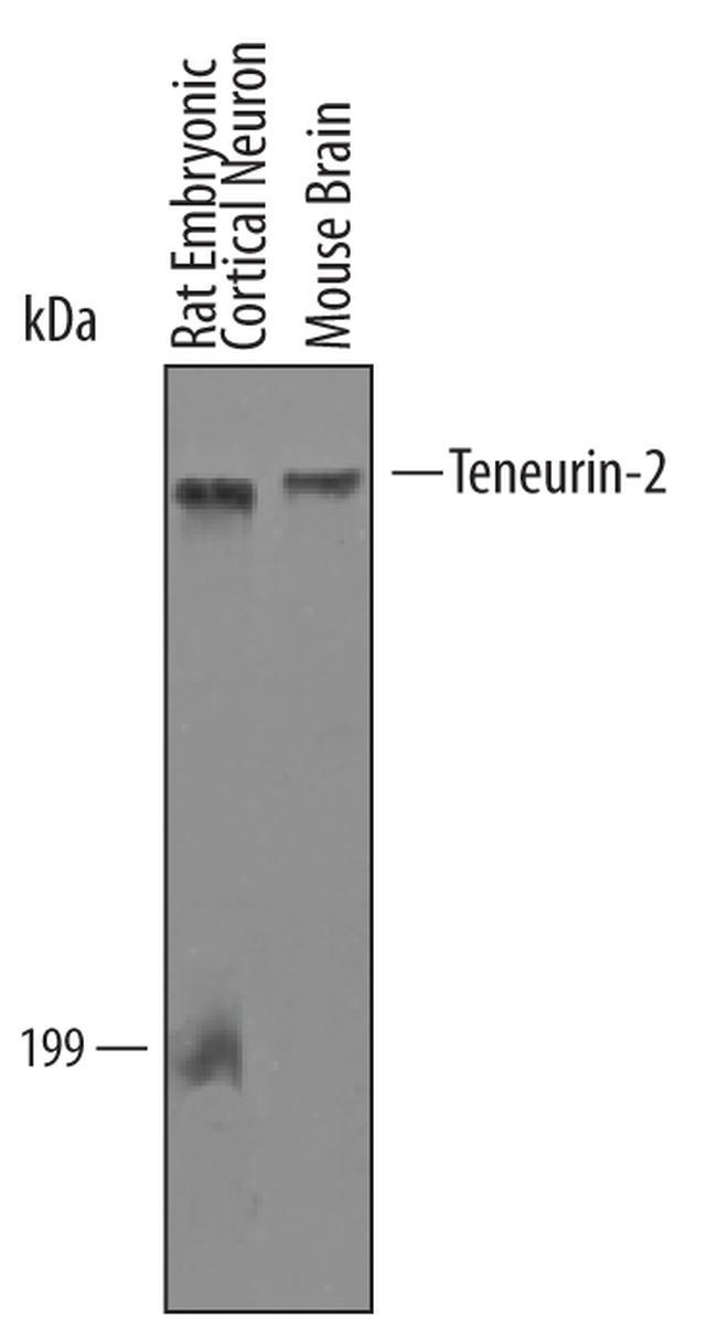
- Western blot analysis from lysates of mouse brain tissue and rat embryonic cortical neuron cells. PVDF Membrane was probed with 1 µg/mL of human Teneurin-2 Antigen Affinity-purified Polyclonal Antibody (Product # PA5-47638) followed by HRP-conjugated Anti-Sheep IgG Secondary Antibody. A specific band was detected for Teneurin-2 at approximately 300 kDa (as indicated). This experiment was conducted under reducing conditions.
- Submitted by
- Invitrogen Antibodies (provider)
- Main image
- Experimental details
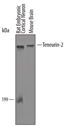
- Western blot analysis of TENM2 in mouse brain tissue and rat embryonic cortical neuron cells. Samples were incubated in TENM2 polyclonal antibody (Product # PA5-47638) using a dilution of 1 µg/mL followed by a HRP-conjugated Anti-Sheep IgG secondary antibody. A specific band was detected for Teneurin‚2 at approximately 300 kDa (as indicated). This experiment was conducted under reducing conditions.

Supportive validation
- Submitted by
- Invitrogen Antibodies (provider)
- Main image

- Experimental details
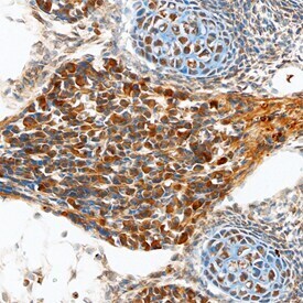
- Immunohistochemical analysis of TENM2 in immersion fixed frozen sections of embryonic mouse brain. Samples were incubated in TENM2 polyclonal antibody (Product # PA5-47638) using a dilution of 10 µg/mL overnight at 4 °C. Tissue was stained using the Anti-Sheep HRP-DAB Cell & Tissue Staining Kit (brown) and counterstained with hematoxylin (blue). Specific staining was localized to neurons in dorsal root ganglia and cells in cartilage primordium.