 Explore
 Validate
 Learn
Learn Western blot
 Immunohistochemistry
Antibody data
- Antibody Data
- Antigen structure
- References [0]
- Comments [0]
- Validations
- Western blot [2]
- Immunocytochemistry [1]
- Immunohistochemistry [9]
Submit
Validation data
Reference
Comment
Report error
- Product number
- HPA036978 - Provider product page

- Provider
- Atlas Antibodies
- Proper citation
- Atlas Antibodies Cat#HPA036978, RRID:AB_10964347
- Product name
- Anti-PGAM5
- Antibody type
- Polyclonal
- Reactivity
- Human
- Host
- Rabbit
- Conjugate
- Unconjugated
- Antigen sequence
NWDRREPLSLINVRKRNVESGEEELASKLDHYKAK
ATRHIFLIRHSQYHVDGSLEKDRTLTPLGREQAEL
TGLRLASLGLKFNKIV- Isotype
- IgG
- Vial size
- 100 µl
- Storage
- Store at +4°C for short term storage. Long time storage is recommended at -20°C.
No comments: Submit comment
Supportive validation
- Submitted by
- Atlas Antibodies (provider)
- Enhanced method
- Genetic validation
- Main image

- Experimental details
- Western blot analysis in RT-4 cells transfected with control siRNA, target specific siRNA probe #1 and #2, using Anti-PGAM5 antibody. Remaining relative intensity is presented. Loading control: Anti-PPIB.

- Submitted by
- Atlas Antibodies (provider)
- Main image

- Experimental details
- Western blot analysis in human cell line A-549.
- Sample type
- HUMAN

Supportive validation
- Submitted by
- Atlas Antibodies (provider)
- Main image

- Experimental details
- Immunofluorescent staining of human cell line MCF7 shows localization to mitochondria.
- Sample type
- HUMAN

Enhanced validation
Supportive validation
- Submitted by
- Atlas Antibodies (provider)
- Enhanced method
- Independent antibody validation
- Main image

- Experimental details
- Immunohistochemical staining of human colon, liver, testis and tonsil using Anti-PGAM5 antibody HPA036978 (A) shows similar protein distribution across tissues to independent antibody HPA036979 (B).

Supportive validation
- Submitted by
- Atlas Antibodies (provider)
- Main image
- Experimental details
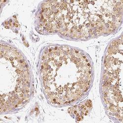
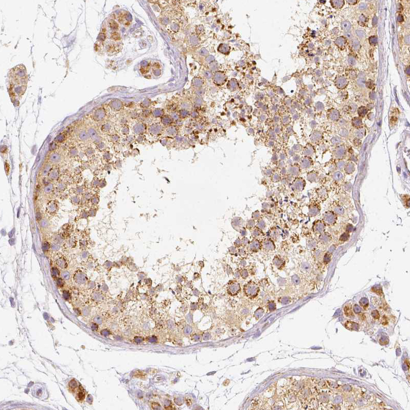
- Immunohistochemical staining of human testis shows moderate to strong granular cytoplasmic positivity in Leydig cells and cells in seminiferous ducts.
- Sample type
- HUMAN

- Submitted by
- Atlas Antibodies (provider)
- Main image

- Experimental details
- Immunohistochemical staining of human kidney shows moderate to strong granular cytoplasmic positivity in cells in tubules.
- Sample type
- HUMAN

- Submitted by
- Atlas Antibodies (provider)
- Main image
- Experimental details
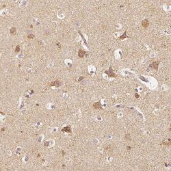
- Immunohistochemical staining of human cerebral cortex shows moderate granular cytoplasmic positivity in neuronal cells.
- Sample type
- HUMAN

- Submitted by
- Atlas Antibodies (provider)
- Main image

- Experimental details
- Immunohistochemical staining of human tonsil shows moderate to strong granular cytoplasmic positivity in lymphoid cells.
- Sample type
- HUMAN

- Submitted by
- Atlas Antibodies (provider)
- Main image

- Experimental details
- Immunohistochemical staining of human liver shows moderate granular cytoplasmic positivity in hepatocytes.
- Sample type
- HUMAN

- Submitted by
- Atlas Antibodies (provider)
- Main image

- Experimental details
- Immunohistochemical staining of human testis shows moderate granular cytoplasmic positivity in cells in seminiferous ducts.
- Sample type
- HUMAN
- Submitted by
- Atlas Antibodies (provider)
- Main image

- Experimental details
- Immunohistochemical staining of human tonsil shows moderate granular cytoplasmic positivity in germinal center cells.
- Sample type
- HUMAN

- Submitted by
- Atlas Antibodies (provider)
- Main image

- Experimental details
- Immunohistochemical staining of human colon shows moderate granular cytoplasmic positivity in glandular cells.
- Sample type
- HUMAN
