Explore
 Validate
 Learn
Learn Western blot
Antibody data
- Antibody Data
- Antigen structure
- References [1]
- Comments [0]
- Validations
- Western blot [1]
- Other assay [1]
Submit
Validation data
Reference
Comment
Report error
- Product number
- PA5-46624 - Provider product page

- Provider
- Invitrogen Antibodies
- Product name
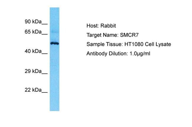
- SMCR7 Polyclonal Antibody
- Antibody type
- Polyclonal
- Antigen
- Synthetic peptide
- Description
- Peptide sequence: FSQKRGKRRS DEGLGSMVDF LLANARLVLG VGGAAVLGIA TLAVKRFIDR
- Concentration
- 0.5 mg/mL
Submitted references HPV/E7 induces chemotherapy-mediated tumor suppression by ceramide-dependent mitophagy.
Thomas RJ, Oleinik N, Panneer Selvam S, Vaena SG, Dany M, Nganga RN, Depalma R, Baron KD, Kim J, Szulc ZM, Ogretmen B
EMBO molecular medicine 2017 Aug;9(8):1030-1051
EMBO molecular medicine 2017 Aug;9(8):1030-1051
No comments: Submit comment
Supportive validation
- Submitted by
- Invitrogen Antibodies (provider)
- Main image

- Experimental details
- Western blot analysis of human HT1080 cell lysate using an anti-SMCR7 polyclonal antibody (Product # PA5-46624).
Supportive validation
- Submitted by
- Invitrogen Antibodies (provider)
- Main image

- Experimental details
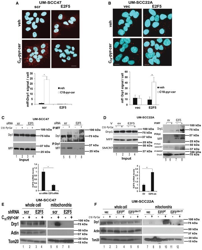
- Figure 9 E2F5 enhances Drp1 translocation to mitochondria and association with MFF to induce HPV-E7/ceramide-dependent mitophagy Effects of stable knockdown of E2F5 using shRNA on DRP1-MFF association with/without C 18 -pyr-cer (10 muM, 1 h) were measured by PLA (scale bars represent 100 mum) using anti-DRP1 and anti-MFF antibodies compared to Scr-shRNA-transfected UM-SCC-47 controls. PLA fluorescence images were quantified using the PLA software as described by the manufacturer. Data are means +- SD from three independent experiments, analyzed by unpaired Student's t -test ( n = 3, * P = 0.0042). HPV(-) UM-SCC-22A cells transiently transfected with exogenous E2F5 or empty vector (vec) were used to measure the association between Drp1 and MFF in the absence/presence of C 18 -pyr-cer (10 muM, 1 h) by PLA (scale bars represent 100 mum) using anti-DRP1 and anti-MFF antibodies. PLA fluorescence images were quantified using the PLA software as described by the manufacturer. Data are means +- SD from three independent experiments, analyzed by unpaired Student's t -test ( n = 3, * P = 0.0051). Effects of C 18 -pyr-cer (10 muM, 1 h) on Drp1-MFF interaction in the absence/presence of E2F5 knockdown using shRNA (versus Scr-shRNA) were measured by immunoprecipitation followed by Western blotting using anti-Drp1 and anti-MFF antibodies (right panel). Equal immunoprecipitation of Drp1 or MFF was confirmed by Western blotting (left panel, input). Blots represent three independent studies