Explore
 Validate
 Learn
Learn Western blot
Antibody data
- Antibody Data
- Antigen structure
- References [0]
- Comments [0]
- Validations
- Western blot [4]
- Immunocytochemistry [2]
- Immunohistochemistry [3]
- Flow cytometry [1]
Submit
Validation data
Reference
Comment
Report error
- Product number
- NBP2-29984 - Provider product page

- Provider
- Novus Biologicals
- Product name
- Rabbit Polyclonal BCKDHB Antibody
- Antibody type
- Polyclonal
- Description
- Protein A purified.
- Reactivity
- Human
- Host
- Rabbit
- Isotype
- IgG
- Vial size
- 0.4 ml
- Concentration
- 0.45 mg/ml
- Storage
- Store at 4C short term. Aliquot and store at -20C long term. Avoid freeze-thaw cycles.
No comments: Submit comment
Supportive validation
- Submitted by
- Novus Biologicals (provider)
- Main image
- Experimental details
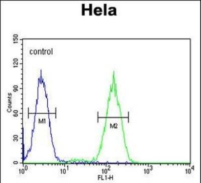
- Western Blot: BCKDHB Antibody [NBP2-29984] - Flow cytometric analysis of Hela cells (right histogram) compared to a negative control cell (left histogram).FITC-conjugated goat-anti-rabbit secondary antibodies were used for the analysis.

- Submitted by
- Novus Biologicals (provider)
- Main image
- Experimental details
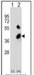
- Western Blot: BCKDHB Antibody [NBP2-29984] - Western blot analysis of BCKDHB (arrow) using rabbit polyclonal (N-term) (NBP2-29984). 293 cell lysates (2 ug/lane) either nontransfected (Lane 1) or transiently transfected (Lane 2) with the BCKDHB gene.

- Submitted by
- Novus Biologicals (provider)
- Main image

- Experimental details
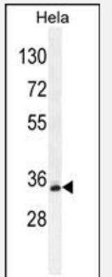
- Western Blot: BCKDHB Antibody [NBP2-29984] - Western blot analysis in Hela cell line lysates (35ug/lane).This demonstrates the BCKDHB antibody detected the BCKDHB protein (arrow).
- Submitted by
- Novus Biologicals (provider)
- Main image
- Experimental details
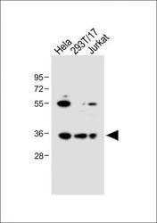
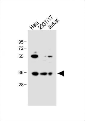
- Western Blot: BCKDHB Antibody (RB20925) [NBP2-29984] - All lanes : Anti-BCKDHB Antibody (N-term). Lane 1: Hela whole cell lysate; Lane 2: 293T/17 whole cell lysate; Lane 3: Jurkat whole cell lysate. Lysates/proteins at 20 ug per lane. Secondary Goat Anti-Rabbit IgG, (H+L), Peroxidase conjugated. Predicted band size : 43 kDa. Blocking/Dilution buffer: 5% NFDM/TBST.
Supportive validation
- Submitted by
- Novus Biologicals (provider)
- Main image
- Experimental details
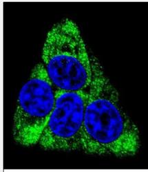
- Immunocytochemistry/Immunofluorescence: BCKDHB Antibody [NBP2-29984] - Confocal immunofluorescent analysis of (N-term)(NBP2-29984) with Hela cell followed by Alexa Fluor 488-conjugated goat anti-rabbit lgG (green). DAPI was used to stain the cell nuclear (blue).

- Submitted by
- Novus Biologicals (provider)
- Main image

- Experimental details
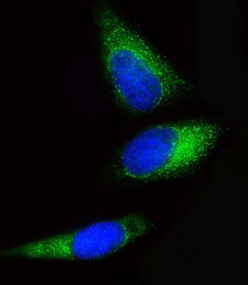
- Immunofluorescence: BCKDHB Antibody (RB20925) [NBP2-29984] - Immunofluorescent analysis of 4% paraformaldehyde-fixed, 0. 1% Triton X-100 permeabilized Hela cells labeling BCKDHB with NBP2-29984, followed by Dylight(R) 488-conjugated goat anti-Rabbit IgG secondary antibody (green). Immunofluorescence image showing Cytoplasm staining on Hela cell line. The nuclear counter stain is DAPI (blue).
Supportive validation
- Submitted by
- Novus Biologicals (provider)
- Main image
- Experimental details
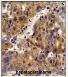
- Immunohistochemistry: BCKDHB Antibody [NBP2-29984] - Immunohistochemistry analysis in formalin fixed and paraffin embedded human hepatocarcinoma followed by peroxidase conjugation of the secondary antibody and DAB staining. This data demonstrates the use of the (N-term) for immunohistochemistry. Clinical relevance has not been evaluated.

- Submitted by
- Novus Biologicals (provider)
- Main image
- Experimental details
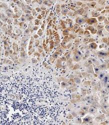
- Immunohistochemistry-Paraffin: BCKDHB Antibody (RB20925) [NBP2-29984] - Immunohistochemical analysis of paraffin-embedded Human hepatocarcinoma tissue using NBP2-29984 performed on the Leica® BOND RXm. Tissue was fixed with formaldehyde at room temperature, antigen retrieval was by heat mediation with a EDTA buffer (pH9. 0). Samples were incubated with primary antibody for 1 hours at RT. A undiluted biotinylated CRF Anti-Polyvalent HRP Polymer antibody was used as the secondary antibody.

- Submitted by
- Novus Biologicals (provider)
- Main image

- Experimental details
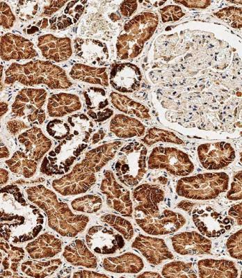
- Immunohistochemistry-Paraffin: BCKDHB Antibody (RB20925) [NBP2-29984] - Immunohistochemical analysis of paraffin-embedded Human kidney tissue using NBP2-29984 performed on the Leica® BOND RXm. Tissue was fixed with formaldehyde at room temperature, antigen retrieval was by heat mediation with a EDTA buffer (pH9. 0). Samples were incubated with primary antibody for 1 hours at RT. A undiluted biotinylated CRF Anti-Polyvalent HRP Polymer antibody was used as the secondary antibody.
Supportive validation
- Submitted by
- Novus Biologicals (provider)
- Main image
- Experimental details
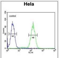
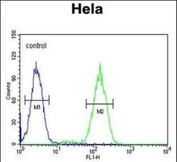
- Flow Cytometry: BCKDHB Antibody (RB20925) [NBP2-29984] - BCKDHB Antibody (N-term) (NBP2-29984) flow cytometric analysis of Hela cells (right histogram) compared to a negative control cell (left histogram).FITC-conjugated goat-anti-rabbit secondary antibodies were used for the analysis.