 Explore
 Validate
 Learn
Learn Immunocytochemistry
Antibody data
- Antibody Data
- Antigen structure
- References [1]
- Comments [0]
- Validations
- Immunocytochemistry [1]
- Immunohistochemistry [3]
- Other assay [1]
Submit
Validation data
Reference
Comment
Report error
- Product number
- 50-6525-82 - Provider product page

- Provider
- Invitrogen Antibodies
- Product name
- Synaptophysin Monoclonal Antibody (EP10), eFluor™ 660, eBioscience™
- Antibody type
- Monoclonal
- Antigen
- Other
- Description
- Description: This EP10 monoclonal antibody reacts with human Synaptophysin, which is also known as Major Synaptic Vesicle Protein p38. This 38-kDa integral membrane protein is expressed on neuronal presynaptic vesicles in the brain, spinal cord, retina, neuromuscular junctions, and adrenal medulla. Synaptophysin is also expressed in neuroendocrine tumors and neuroblastomas. Synaptophysin has been reported to play a role in synaptic vesicle trafficking. The EP10 antibody recognizes human and hamster synaptophysin but does not recognize mouse and rat synaptophysin. Applications Reported: This EP10 antibody has been reported for use in immunohistochemical staining, and immunocytochemistry. Applications Tested: This EP10 antibody has been tested by immunohistochemistry on FFPE human tissue using low pH antigen retrieval at less than or equal to 10 µg/mL. It is recommended that the antibody be carefully titrated for optimal performance in the assay of interest. eFluor® 660 is a replacement for Alexa Fluor® 647. eFluor® 660 emits at 659 nm and is excited with the red laser (633 nm). Please make sure that your instrument is capable of detecting this fluorochome. Excitation: 633-647 nm; Emission: 668 nm; Laser: Red Laser. Filtration: 0.2 µm post-manufacturing filtered.
- Reactivity
- Human, Hamster
- Host
- Mouse
- Isotype
- IgG
- Antibody clone number
- EP10
- Vial size
- 100 µg
- Concentration
- 0.2 mg/mL
- Storage
- 4° C, store in dark, DO NOT FREEZE!
Submitted references Cerebral organoids at the air-liquid interface generate diverse nerve tracts with functional output.
Giandomenico SL, Mierau SB, Gibbons GM, Wenger LMD, Masullo L, Sit T, Sutcliffe M, Boulanger J, Tripodi M, Derivery E, Paulsen O, Lakatos A, Lancaster MA
Nature neuroscience 2019 Apr;22(4):669-679
Nature neuroscience 2019 Apr;22(4):669-679
No comments: Submit comment
Supportive validation
- Submitted by
- Invitrogen Antibodies (provider)
- Main image

- Experimental details
- Immunofluorescence analysis of Synaptophysin was performed using SH-SY5Y cells differentiated into neurons. The cells were fixed with 4% paraformaldehyde for 10 minutes, permeabilized with 0.1% Triton™ X-100 for 15 minutes, and blocked with 2% BSA for 1 hour at room temperature. The cells were labeled with Synaptophysin Monoclonal Antibody (EP10), eFluor 660, eBioscience™ (Product # 50-6525-82) at 5 µg/mL in 0.1% BSA, incubated at 4 degree celsius overnight (Panel a: Red). Nuclei (Panel b: Blue) were stained with ProLong™ Diamond Antifade Mountant with DAPI (Product # P36962). F-actin (Panel c: Green) was stained with Rhodamine Phalloidin (Product # R415, 1:300 dilution). Panel d represents the merged image showing cytoplasmic localization. Panel e represents undifferentiated SH-SY5Y cells, showing lower levels of Synaptophysin expression. Panel f represents control cells with isotype control antibody to assess background. The images were captured at 60X magnification.

Supportive validation
- Submitted by
- Invitrogen Antibodies (provider)
- Main image

- Experimental details
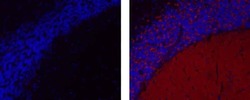
- Immunohistochemistry of formalin-fixed paraffin embedded human cerebellum using 10 µg/mL Mouse IgG1 K Isotype Control eFluor® 660 (left) or 10 µg/mL Anti-Human Synaptophysin eFluor® 660 (right). Nuclei are stained with DAPI.

- Submitted by
- Invitrogen Antibodies (provider)
- Main image

- Experimental details
- Immunohistochemistry of formalin-fixed paraffin embedded human cerebellum using 10 µg/mL Mouse IgG1 K Isotype Control eFluor® 660 (left) or 10 µg/mL Anti-Human Synaptophysin eFluor® 660 (right). Nuclei are stained with DAPI.

- Submitted by
- Invitrogen Antibodies (provider)
- Main image
- Experimental details
- Immunohistochemistry of formalin-fixed paraffin embedded human cerebellum using 10 µg/mL Mouse IgG1 K Isotype Control eFluor® 660 (left) or 10 µg/mL Anti-Human Synaptophysin eFluor® 660 (right). Nuclei are stained with DAPI.

Supportive validation
- Submitted by
- Invitrogen Antibodies (provider)
- Main image

- Experimental details
- Figure 2 ALI-CO cultures exhibit mature neuronal morphology and function a. Sparse labeling in an ALI-CO at 37 days at the ALI (80 days total) by Sendai-virus encoding emGFP (white) reveals radially aligned neurons (NEUROD2, blue) with complex dendritic architectures and pyramidal morphologies (arrowheads) within the aligned cortical plate (bracket). All cells contain fFusionRed (red) to visualize the overall tissue morphology. b. Higher magnification of a maximum intensity projection of a single emGFP (green) labeled neuron (NEUROD2 + , magenta) displaying typical pyramidal morphology with primary dendrite (arrow) and basal dendrites (arrowheads). Synaptophysin (white) reveals extensive synaptic staining throughout. c. Electroporation of membrane targeted farnesylated GFP (fGFP, green) reveals complex dendritic architecture of radially aligned pyramidal neurons (arrows) in a maximum intensity projection with evident dendritic spines (arrowheads, inset) in an ALI-CO at 51 days at the ALI (143 days total) d. Sparse labeling of a 1-year ALI-CO (90 + 275 days ALI) with Sendai-emGFP labels several individual neurons (arrows) with highly complex dendritic architectures and abundant dendritic spines (arrowheads). Sparse labeling with emGFP and fGFP in a-d were performed on three ALI-COs from three organoids with similar results. e. Two minutes of spontaneous activity recorded from a single electrode of a multi-electrode array (MEA) in an ALI-CO at 54 days at the ALI (117 days total
