 Explore
 Validate
 Learn
Learn Western blot
Antibody data
- Antibody Data
- Antigen structure
- References [1]
- Comments [0]
- Validations
- Western blot [2]
- Immunocytochemistry [1]
Submit
Validation data
Reference
Comment
Report error
- Product number
- AF3206 - Provider product page

- Provider
- R&D Systems
- Product name
- Human/Mouse/Rat Lyn Antibody
- Antibody type
- Polyclonal
- Description
- Antigen Affinity-purified. Detects human, mouse, and rat Lyn in Western blots.
- Reactivity
- Human, Mouse, Rat
- Host
- Goat
- Conjugate
- Unconjugated
- Antigen sequence
P07948- Isotype
- IgG
- Vial size
- 100 ug
- Concentration
- LYOPH
- Storage
- Use a manual defrost freezer and avoid repeated freeze-thaw cycles. 12 months from date of receipt, -20 to -70 °C as supplied. 1 month, 2 to 8 °C under sterile conditions after reconstitution. 6 months, -20 to -70 °C under sterile conditions after reconstitution.
Submitted references Quantitative analysis of membrane remodeling at the phagocytic cup.
Lee WL, Mason D, Schreiber AD, Grinstein S
Molecular biology of the cell 2007 Aug;18(8):2883-92
Molecular biology of the cell 2007 Aug;18(8):2883-92
No comments: Submit comment
Supportive validation
- Submitted by
- R&D Systems (provider)
- Main image
- Experimental details
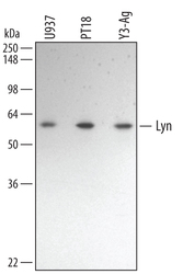
- Detection of Human/Mouse/Rat Lyn by Western Blot. Western blot shows lysates of U937 human histiocytic lymphoma cell line, PT18 mouse mast/basophil cell line, and Y3-Ag rat myeloid cell line. PVDF membrane was probed with 0.5 µg/mL of Goat Anti-Human/Mouse/Rat Lyn Antigen Affinity-purified Polyclonal Antibody (Catalog # AF3206) followed by HRP-conjugated Anti-Goat IgG Secondary Antibody (Catalog # HAF109). A specific band was detected for Lyn at approximately 56 kDa (as indicated). This experiment was conducted under reducing conditions and using Immunoblot Buffer Group 1.
- Submitted by
- R&D Systems (provider)
- Main image
- Experimental details
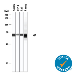
- Detection of Human Lyn by Simple WesternTM. Simple Western lane view shows lysates of Nalm-6 human Pre-B acute lymphocytic leukemia cell line, THP-1 human acute monocytic leukemia cell line, Raji human Burkitt's lymphoma cell line, and Ramos human Burkitt's lymphoma cell line, loaded at 0.2 mg/mL. A specific band was detected for Lyn at approximately 58-60 kDa (as indicated) using 5 µg/mL of Goat Anti-Human/Mouse/Rat Lyn Antigen Affinity-purified Polyclonal Antibody (Catalog # AF3206) followed by 1:50 dilution of HRP-conjugated Anti-Goat IgG Secondary Antibody (Catalog # HAF109). This experiment was conducted under reducing conditions and using the 12-230 kDa separation system.

Supportive validation
- Submitted by
- R&D Systems (provider)
- Main image
- Experimental details
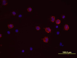
- Lyn in Human PBMCs. Lyn was detected in immersion fixed human peripheral blood mononuclear cells (PBMCs) treated with monensin using Goat Anti-Human/Mouse/Rat Lyn Antigen Affinity-purified Polyclonal Antibody (Catalog # AF3206) at 10 µg/mL for 3 hours at room temperature. Cells were stained using the NorthernLights™ 557-conjugated Anti-Goat IgG Secondary Antibody (red; Catalog # NL001) and counterstained with DAPI (blue). View our protocol for Fluorescent ICC Staining of Non-adherent Cells.
