 Explore
 Validate
 Learn
Learn Immunohistochemistry
Antibody data
- Antibody Data
- Antigen structure
- References [1]
- Comments [0]
- Validations
- Immunohistochemistry [7]
Submit
Validation data
Reference
Comment
Report error
- Product number
- HPA037002 - Provider product page

- Provider
- Atlas Antibodies
- Proper citation
- Atlas Antibodies Cat#HPA037002, RRID:AB_10672383
- Product name
- Anti-BANK1
- Antibody type
- Polyclonal
- Reactivity
- Human
- Host
- Rabbit
- Conjugate
- Unconjugated
- Antigen sequence
RHGHKELKKIFEDFSIQEIDINNEQENDYEEDIAS
FSTYIPSTQNPAFHHESRKTYGQSADGAEANEMEG
EGKQNGSGMETKHSPL- Isotype
- IgG
- Vial size
- 100 µl
- Storage
- Store at +4°C for short term storage. Long time storage is recommended at -20°C.
Submitted references BANK1 and BLK act through phospholipase C gamma 2 in B-cell signaling.
Bernal-Quirós M, Wu YY, Alarcón-Riquelme ME, Castillejo-López C
PloS one 2013;8(3):e59842
PloS one 2013;8(3):e59842
No comments: Submit comment
Enhanced validation
Supportive validation
- Submitted by
- Atlas Antibodies (provider)
- Enhanced method
- Orthogonal validation
- Main image

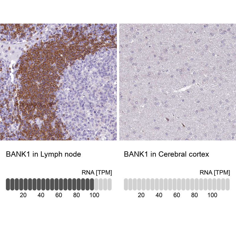
- Experimental details
- Immunohistochemistry analysis in human lymph node and cerebral cortex tissues using HPA037002 antibody. Corresponding BANK1 RNA-seq data are presented for the same tissues.
- Sample type
- HUMAN
Supportive validation
- Submitted by
- Atlas Antibodies (provider)
- Main image

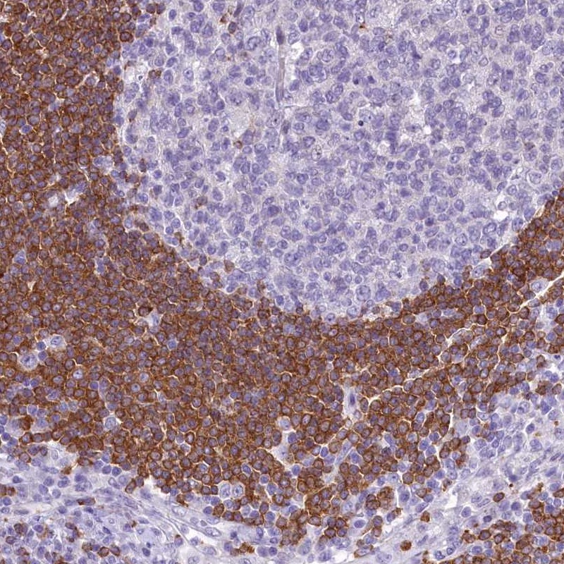
- Experimental details
- Immunohistochemical staining of human lymph node shows high expression.
- Sample type
- HUMAN
- Submitted by
- Atlas Antibodies (provider)
- Main image

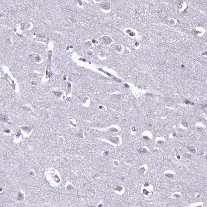
- Experimental details
- Immunohistochemical staining of human cerebral cortex shows low expression as expected.
- Sample type
- HUMAN
- Submitted by
- Atlas Antibodies (provider)
- Main image
- Experimental details
- Immunohistochemical staining of human tonsil shows moderate to strong cytoplasmic positivity in germinal center cells.
- Sample type
- HUMAN

- Submitted by
- Atlas Antibodies (provider)
- Main image

- Experimental details
- Immunohistochemical staining of human fallopian tube shows weak to moderate cytoplasmic positivity in glandular cells.
- Sample type
- HUMAN

- Submitted by
- Atlas Antibodies (provider)
- Main image

- Experimental details
- Immunohistochemical staining of human cerebral cortex shows no positivity in neurons as expected.
- Sample type
- HUMAN

- Submitted by
- Atlas Antibodies (provider)
- Main image

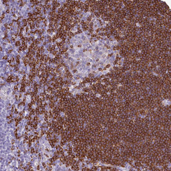
- Experimental details
- Immunohistochemical staining of human lymph node shows moderate to strong cytoplasmic positivity in germinal center cells.
- Sample type
- HUMAN
