Explore
 Validate
 Learn
Learn Immunocytochemistry
Antibody data
- Antibody Data
- Antigen structure
- References [1]
- Comments [0]
- Validations
- Immunocytochemistry [1]
- Other assay [2]
Submit
Validation data
Reference
Comment
Report error
- Product number
- PA5-83256 - Provider product page

- Provider
- Invitrogen Antibodies
- Product name
- HFM1 Polyclonal Antibody
- Antibody type
- Polyclonal
- Antigen
- Recombinant full-length protein
- Description
- Immunogen sequence: SSVPPVKRLK IQMNKSQSVD LKEFGFTPKP SLPSISRSEY LNISELPIME QWDQPEIYGK VRQEPSEYQD KEVLNVNFEL GNEVWDD
- Reactivity
- Human
- Host
- Rabbit
- Isotype
- IgG
- Vial size
- 100 µL
- Concentration
- 0.2 mg/mL
- Storage
- Store at 4°C short term. For long term storage, store at -20°C, avoiding freeze/thaw cycles.
Submitted references Hfm1 participates in Golgi-associated spindle assembly and division in mouse oocyte meiosis.
Wang H, Zhong C, Yang R, Yin Y, Tan R, Gao L, Gao C, Cui Y, Pu D, Wu J
Cell death & disease 2020 Jun 30;11(6):490
Cell death & disease 2020 Jun 30;11(6):490
No comments: Submit comment
Supportive validation
- Submitted by
- Invitrogen Antibodies (provider)
- Main image
- Experimental details
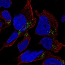
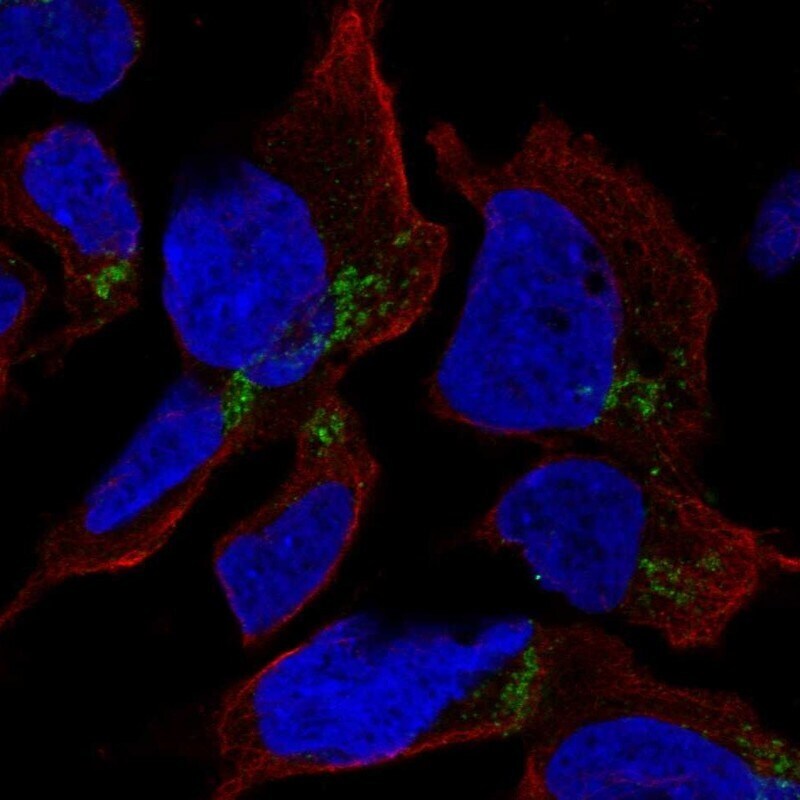
- Immunofluorescent analysis of HFM1 in AF22 cells using a HFM1 polyclonal antibody (Product # PA5-83256). The analysis shows localization to the Golgi apparatus.
Supportive validation
- Submitted by
- Invitrogen Antibodies (provider)
- Main image
- Experimental details
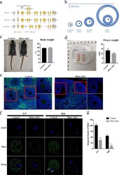
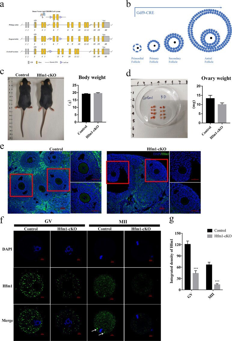
- Fig. 1 Oocyte-specific Knockout of Hfm1 in mice. a Engineered a conditional floxed allele for Hfm1 and a Cre-mediated recombination to delete exons 6 and 7. b Schematic illustration of Gdf9-Cre-mediated Hfm1 knockout in oocytes from primordial follicle stage c Photograph of whole bodies and measured the body weight of Control and Hfm1-cKO mice (2 months old) ( n = 4). d Photograph of a pair of ovaries and measured the ovaries weight of Control and Hfm1-cKO mice (2-month-old) ( n = 4). e Immunofluorescence staining of Hfm1 in ovaries of Control and Hfm1-cKO mice (2-month-old). f Immunofluorescence showed expression levels and subcellular localization of Hfm1 (green) in GV and MII oocytes of Control and Hfm1-cKO mice (2-month-old). DAPI are co-stained to visualize DNA (blue). White arrows point to spindle pole in oocytes. GV, Germinal Vesicle; MII, metaphase of meiosis II. g Quantification of the Hfm1 staining ( n = 10). *** P < 0.01 by two-tailed Student's t tests. Data represent the mean +- SEM.
- Submitted by
- Invitrogen Antibodies (provider)
- Main image
- Experimental details
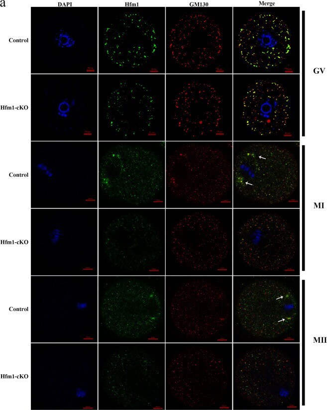
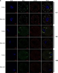
- Fig. 4 Hfm1 functions in Golgi-associated spindle assembly and division. a Double immunofluorescent staining of Hfm1 (green) and GM130 (red) in GV, MI and MII stage. DAPI are co-stained to visualize DNA (blue). White arrows point to spindle pole in oocytes. GV germinal vesicle, MI metaphase of meiosis I, MII metaphase of meiosis II.