Explore
 Validate
 Learn
Learn Western blot
Antibody data
- Antibody Data
- Antigen structure
- References [0]
- Comments [0]
- Validations
- Western blot [1]
- Immunocytochemistry [2]
- Immunohistochemistry [1]
Submit
Validation data
Reference
Comment
Report error
- Product number
- PA5-58249 - Provider product page

- Provider
- Invitrogen Antibodies
- Product name
- FAM13A Polyclonal Antibody
- Antibody type
- Polyclonal
- Antigen
- Recombinant full-length protein
- Description
- Immunogen sequence: ESGTLSASSA TSARQRRRQS KEQDEVRHGR DKGLINKENT PSGFNHLDDC ILNTQEVEKV HKNTFGCAGE RS
- Concentration
- 0.1 mg/mL
No comments: Submit comment
Supportive validation
- Submitted by
- Invitrogen Antibodies (provider)
- Main image
- Experimental details
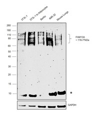
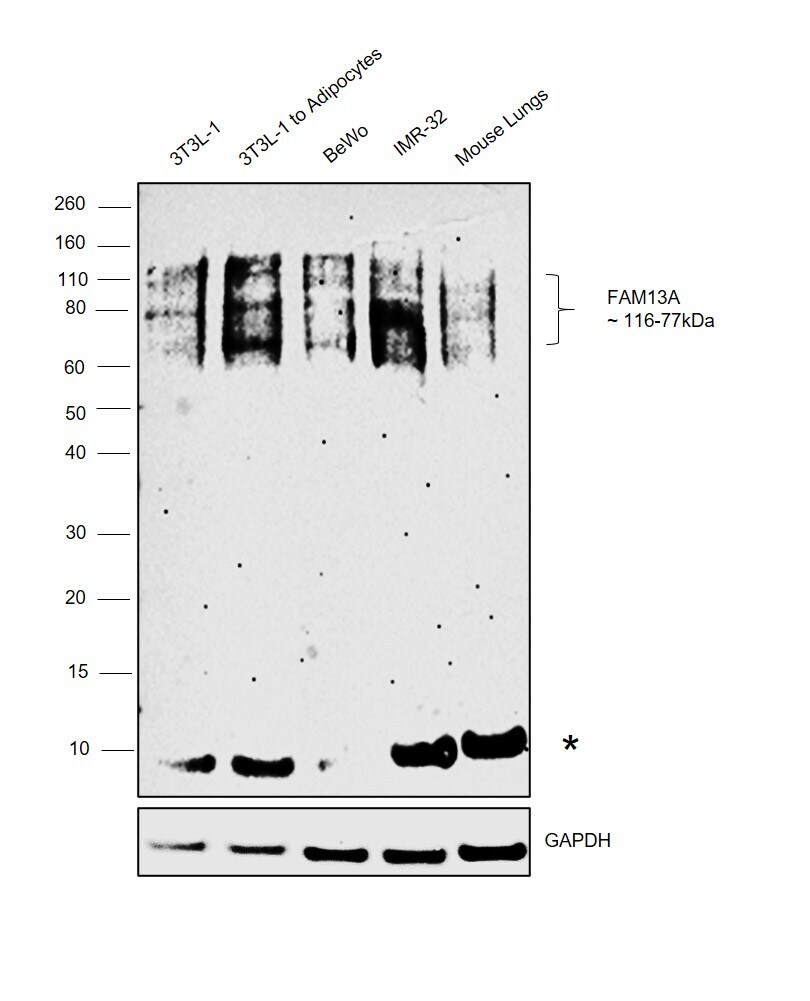
- Western blot was performed using Anti-FAM13A Polyclonal Antibody (Product # PA5-58249) and a 116-77kDa band corresponding to Protein FAM13A was observed across the cell lines and tissues tested and increased upon 3T3L-1 differentiation to adipocytes along with an uncharacterized band around 10kDa. Whole cell extracts (30 µg lysate) of 3T3-L1 (Lane 1), 3T3-L1 differentiated to adipocytes (Lane 2), BeWo (Lane 3), IMR-32 (Lane 4) and tissue extracts of Mouse Lung (Lane 5) were electrophoresed using NuPAGE™ 4-12% Bis-Tris Protein Gel (Product # NP0321BOX). Resolved proteins were then transferred onto a nitrocellulose membrane (Product # LC2001) by iBlot® 2 Dry Blotting System (Product # IB21001). The blot was probed with the primary antibody at 1:1000 dilution and detected by chemiluminescence with Goat anti-Rabbit IgG (H+L) Superclonal™ Recombinant Secondary Antibody, HRP (Product # A27036, 1:4000 dilution) using the iBright FL 1000 (Product # A32752). Chemiluminescent detection was performed using Novex® ECL Chemiluminescent Substrate Reagent Kit (Product # WP20005).
Supportive validation
- Submitted by
- Invitrogen Antibodies (provider)
- Main image

- Experimental details
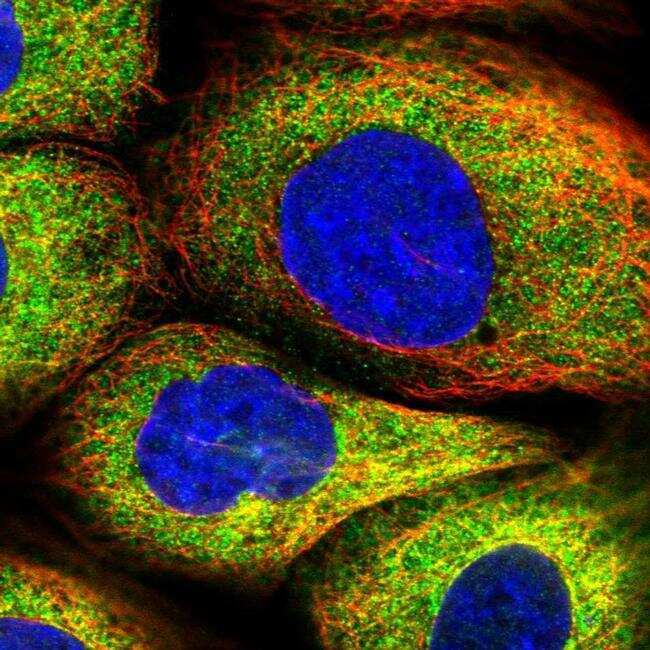
- Immunofluorescent staining of FAM13A in human cell line A-431 shows positivity in cytoplasm. Samples were probed using a FAM13A Polyclonal Antibody (Product # PA5-58249).
- Submitted by
- Invitrogen Antibodies (provider)
- Main image
- Experimental details
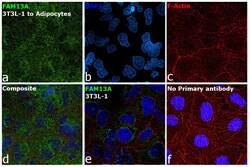
- Immunofluorescence analysis of Protein FAM13A was performed using 70% confluent log phase 3T3-L1 cells differentiated to Adipocytes. The cells were fixed with 4% paraformaldehyde for 15 minutes, permeabilized with 0.1% Triton™ X-100 for 10 minutes, and blocked with 2% BSA for 45 minutes at room temperature. The cells were labeled with FAM13A Polyclonal Antibody (Product # PA5-58249) at 1:200 dilution in 0.1% BSA, incubated at 4 degree celsius overnight and then labeled with Goat anti-Rabbit IgG (H+L) Superclonal™ Recombinant Secondary Antibody, Alexa Fluor® 488 conjugate (Product # A27034), (1:2000 dilution), for 45 minutes at room temperature (Panel a: Green). Nuclei (Panel b:Blue) were stained with ProLong™ Diamond Antifade Mountant with DAPI (Product # P36962). F-actin (Panel c: Red) was stained with Rhodamine Phalloidin (Product # R415, 1:300 dilution). Panel d represents the merged image showing cytoplasmic localization. Panel e represents undifferentiated 3T3L-1 cells showing lower expression of FAM13A. Panel f represents control 3T3L-1 cells with no primary antibody to assess background. The images were captured at 60X magnification.
Supportive validation
- Submitted by
- Invitrogen Antibodies (provider)
- Main image
- Experimental details
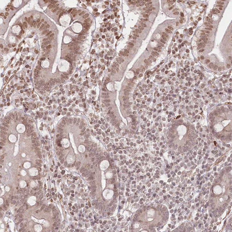
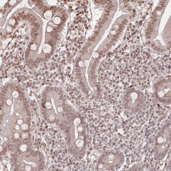
- Immunohistochemical staining of FAM13A in human duodenum using a FAM13A Polyclonal Antibody (Product # PA5-58249) shows nuclear positivity in glandular cells.