 Explore
 Validate
 Learn
Learn Western blot
Antibody data
- Antibody Data
- Antigen structure
- References [1]
- Comments [0]
- Validations
- Western blot [2]
- Immunohistochemistry [1]
- Other assay [1]
Submit
Validation data
Reference
Comment
Report error
- Product number
- PA5-21157 - Provider product page

- Provider
- Invitrogen Antibodies
- Product name
- TEKT5 Polyclonal Antibody
- Antibody type
- Polyclonal
- Antigen
- Synthetic peptide
- Description
- A suggested positive control is NIH-3T3 cell lysate.
- Concentration
- 1 mg/mL
Submitted references Comprehensive Analysis of Mouse Cancer/Testis Antigen Functions in Cancer Cells and Roles of TEKT5 in Cancer Cells and Testicular Germ Cells.
Aoki N, Matsui Y
Molecular and cellular biology 2019 Sep 1;39(17)
Molecular and cellular biology 2019 Sep 1;39(17)
No comments: Submit comment
Supportive validation
- Submitted by
- Invitrogen Antibodies (provider)
- Main image

- Experimental details
- Western blot analysis of 3T3 cell lysate using a TEKT5 polyclonal antibody (Product # PA5-21157) at (A) 0.25 and (B) 0.5 µg/mL.

- Submitted by
- Invitrogen Antibodies (provider)
- Main image

- Experimental details
- Western Blot analysis of TEKT5 in 3T3 cell lysate with TEKT5 Polyclonal Antibody (Product # PA5-21157) at (A) 0.25 and (B) 0.5 µg/mL.

Supportive validation
- Submitted by
- Invitrogen Antibodies (provider)
- Main image

- Experimental details
- Immunohistochemistry of TEKT5 in rat testis tissue with TEKT5 Polyclonal Antibody (Product # PA5-21157) at 5 µg/mL.

Supportive validation
- Submitted by
- Invitrogen Antibodies (provider)
- Main image
- Experimental details
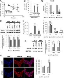
- Repression of G1-S transition of cell cycle and cell survival and increased nuclear localization of SMAD3 by Tekt5-KD in OV3121 and MH134-TC cells. (A and B) Changes in viability of an ovarian cancer cell line, OV3121 (A), and of a liver cancer cell line, MH134-TC (B), by two different siRNA corresponding to Tekt5 (siTekt5#1 and siTekt5#2). siCTL, AllStars negative control siRNA. MH134-TC cells were assayed at 72h after transfection with the siRNAs. Cell viability was determined by MTS assay. (C and D) Decreased accumulation of Tekt5 mRNA as determined by RT-qPCR (C) and of TEKT5 protein as determined by Western blotting (D, upper) after Tekt5-KD in OV3121 cells. For the Western blotting, the signal intensity of TEKT5 was quantified and then normalized against that of the housekeeping protein GAPDH (D, lower). (E and F) Ratios of active caspase-3-positive apoptotic cells (E) and of cells in each phase of the cell cycle (F) at 48 h after transfection of OV3121 cells with Tekt5 siRNA (siTekt5#1), as determined by flow cytometry. (G) Accumulation of p27kip 24 and 48h after transfection of OV3121 cells with Tekt5 siRNA, as determined by Western blotting (left). The signal intensity of p27kip was quantified and then normalized against that of GAPDH (right). (H) Localization of SMAD3 24 and 48 h after transfection of OV3121 cells with Tekt5 siRNA, as determined by immunostaining (left). SMAD3 accumulation is shown in red at 48 h in the micrographs. Nuclear staining by DAPI (blue)
