Explore
 Validate
 Learn
Learn Western blot
 ELISA
ELISAAntibody data
- Antibody Data
- Antigen structure
- References [4]
- Comments [0]
- Validations
- Western blot [1]
- Immunocytochemistry [1]
- Immunohistochemistry [1]
- Flow cytometry [1]
Submit
Validation data
Reference
Comment
Report error
- Product number
- ABIN951978 - Provider product page

- Provider
- antibodies-online
- Product name
- anti-Dipeptidyl-Peptidase 3 (DPP3) (C-Term), (AA 590-620) antibody
- Antibody type
- Polyclonal
- Antigen
- KLH conjugated synthetic peptide between 590-620 amino acids from the C-terminal region of Human DPP3
- Description
- Affinity Chromatography on Protein A
- Reactivity
- Human
- Host
- Rabbit
- Epitope
- C-Term,AA 590-620
- Vial size
- 0.4 mL
- Concentration
- 0.25 mg/mL
- Storage
- Store undiluted at 2-8°C for one month or (in aliquots) at -20°C for longer.
- Handling
- Avoid repeated freezing and thawing.
Submitted references Ets-1/Elk-1 is a critical mediator of dipeptidyl-peptidase III transcription in human glioblastoma cells.
Functional tyrosine residue in the active center of human dipeptidyl peptidase III.
Global phosphoproteome analysis on human HepG2 hepatocytes using reversed-phase diagonal LC.
Highly reactive cysteine residues are part of the substrate binding site of mammalian dipeptidyl peptidases III.
Shukla AA, Jain M, Chauhan SS
The FEBS journal 2010 Apr;277(8):1861-75
The FEBS journal 2010 Apr;277(8):1861-75
Functional tyrosine residue in the active center of human dipeptidyl peptidase III.
Salopek-Sondi B, Vukelić B, Spoljarić J, Simaga S, Vujaklija D, Makarević J, Jajcanin N, Abramić M
Biological chemistry 2008 Feb;389(2):163-7
Biological chemistry 2008 Feb;389(2):163-7
Global phosphoproteome analysis on human HepG2 hepatocytes using reversed-phase diagonal LC.
Gevaert K, Staes A, Van Damme J, De Groot S, Hugelier K, Demol H, Martens L, Goethals M, Vandekerckhove J
Proteomics 2005 Sep;5(14):3589-99
Proteomics 2005 Sep;5(14):3589-99
Highly reactive cysteine residues are part of the substrate binding site of mammalian dipeptidyl peptidases III.
Abramić M, Simaga S, Osmak M, Cicin-Sain L, Vukelić B, Vlahovicek K, Dolovcak L
The international journal of biochemistry & cell biology 2004 Mar;36(3):434-46
The international journal of biochemistry & cell biology 2004 Mar;36(3):434-46
No comments: Submit comment
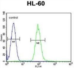
Supportive validation
- Submitted by
- antibodies-online (provider)
- Main image

- Experimental details
- Western blot analysis of DPP3 Antibody (C-term) Cat.-No AP51313PU-N in HL-60 cell line lysates (35ug/lane). This demonstrates the DPP3 antibody detected the DPP3 protein (arrow).

Supportive validation
- Submitted by
- antibodies-online (provider)
- Main image
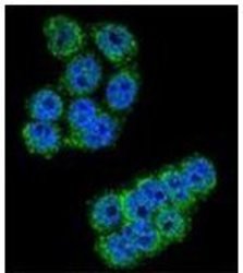
- Experimental details
- Confocal immunofluorescent analysis of DPP3 Antibody (C-term) Cat.-No AP51313PU-N with Hela cell followed by Alexa Fluor 488-conjugated goat anti-rabbit lgG (green). DAPI was used to stain the cell nuclear (blue).

Supportive validation
- Submitted by
- antibodies-online (provider)
- Main image
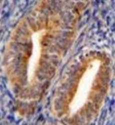
- Experimental details
- Formalin fixed and paraffin embedded human uterus tissue reacted with DPP3 Antibody (C-term) Cat.-No AP51313PU-N followed by peroxidase conjugation of the secondary antibody and DAB staining.

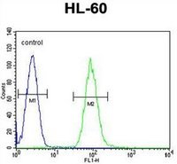
Supportive validation
- Submitted by
- antibodies-online (provider)
- Main image
- Experimental details
- Flow cytometric analysis of HL-60 cells using DPP3 Antibody (C-term) Cat.-No AP51313PU-N (right histogram) compared to a negative control cell (left histogram). FITC-conjugated goat-anti-rabbit secondary antibodies were used for the analysis.