Explore
 Validate
 Learn
LearnPA5-78130
antibody from Invitrogen Antibodies
Targeting: PRKDC
DNA-PKC, DNA-PKcs, DNAPK, DNAPKc, DNPK1, HYRC, HYRC1, p350, p460, XRCC7
 Western blot
Antibody data
- Antibody Data
- Antigen structure
- References [2]
- Comments [0]
- Validations
- Western blot [2]
- Immunocytochemistry [2]
- Chromatin Immunoprecipitation [1]
- Other assay [1]
Submit
Validation data
Reference
Comment
Report error
- Product number
- PA5-78130 - Provider product page

- Provider
- Invitrogen Antibodies
- Product name
- Phospho-DNA-PK (Ser2056) Polyclonal Antibody
- Antibody type
- Polyclonal
- Antigen
- Synthetic peptide
- Description
- Positive Control: 293T, 293T (100 J/m2 UVC treatment)
- Concentration
- 0.76 mg/mL
Submitted references Intrinsic ATR signaling shapes DNA end resection and suppresses toxic DNA-PKcs signaling.
The Ig superfamily protein PTGFRN coordinates survival signaling in glioblastoma multiforme.
Dibitetto D, Sims JR, Ascenção CFR, Feng K, Kim D, Oberly S, Freire R, Smolka MB
NAR cancer 2020 Jun;2(2):zcaa006
NAR cancer 2020 Jun;2(2):zcaa006
The Ig superfamily protein PTGFRN coordinates survival signaling in glioblastoma multiforme.
Aguila B, Morris AB, Spina R, Bar E, Schraner J, Vinkler R, Sohn JW, Welford SM
Cancer letters 2019 Oct 10;462:33-42
Cancer letters 2019 Oct 10;462:33-42
No comments: Submit comment
Supportive validation
- Submitted by
- Invitrogen Antibodies (provider)
- Main image
- Experimental details
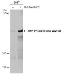
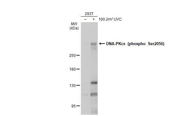
- Western blot analysis of Phospho-DNA-PKcs (Ser2056) in untreated (-) and treated (+) 293T cells using 60 µg of protein. Samples were separated with 5% SDS-PAGE and incubated with Phospho-DNA-PKcs (Ser2056) polyclonal antibody (Product # PA5-78130) using a dilution of 1:1000.
- Submitted by
- Invitrogen Antibodies (provider)
- Main image

- Experimental details
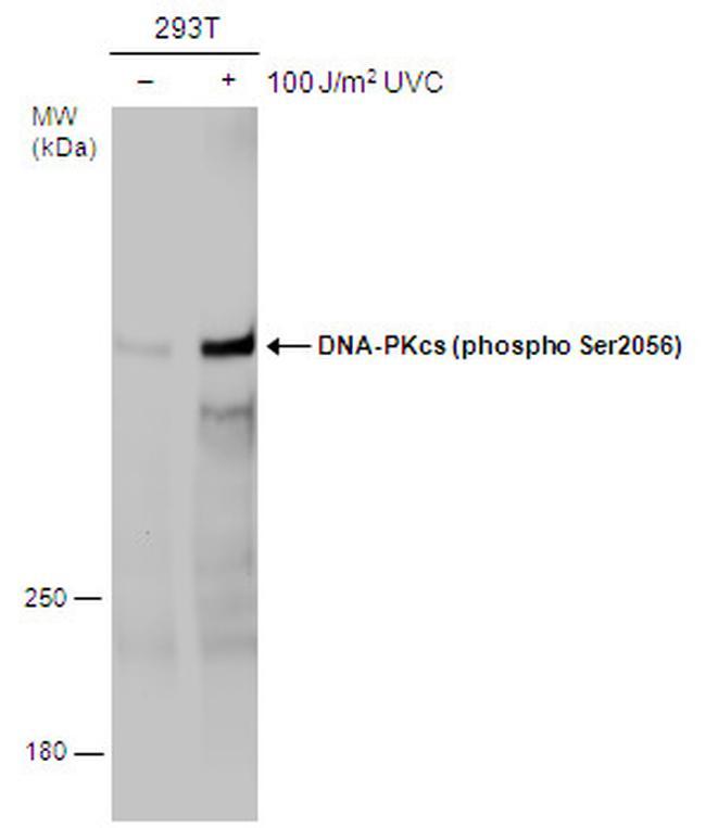
- Western Blot using Phospho-DNA-PK (Ser2056) Polyclonal Antibody (Product # PA5-78130). Untreated (–) and treated (+) 293T whole cell extracts (60 µg) were separated by 5% SDS-PAGE, and the membrane was blotted with Phospho-DNA-PK (Ser2056) Polyclonal Antibody (Product # PA5-78130) diluted at 1:500. The HRP-conjugated anti-rabbit IgG antibody was used to detect the primary antibody.
Supportive validation
- Submitted by
- Invitrogen Antibodies (provider)
- Main image
- Experimental details
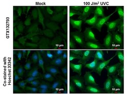
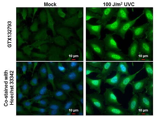
- Immunocytochemistry-Immunofluorescence analysis of Phospho-DNA-PK-(Ser2056) was performed in Mock and treated HeLa cells fixed in 4% paraformaldehyde at RT for 15 min. Green: Phospho-DNA-PK (Ser2056) Polyclonal Antibody (Product # PA5-78130) diluted at 1:500. Blue: Hoechst 33342 staining. Scale bar = 10 µm.
- Submitted by
- Invitrogen Antibodies (provider)
- Main image
- Experimental details
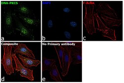
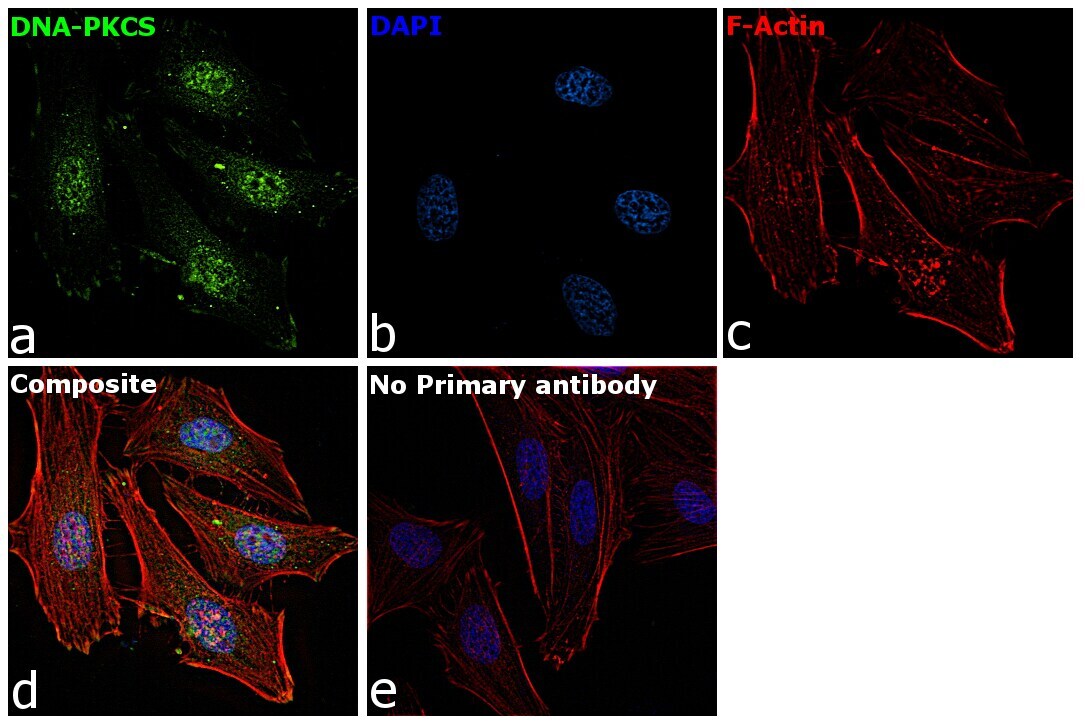
- Immunofluorescence analysis of DNA-PKCS was performed using HeLa cells. The cells were fixed with 4% paraformaldehyde for 10 minutes, permeabilized with 0.1% Triton™ X-100 for 15 minutes, and blocked with 2% BSA for 1 hour at room temperature. The cells were labeled with Phospho-DNA-PK (Ser2056) Polyclonal Antibody (Product # PA5-78130) at 5 µg/mL in 0.1% BSA and incubated overnight at 4 degree and then labeled with Goat anti-Rabbit IgG (H+L) Superclonal™ Recombinant Secondary Antibody, Alexa Fluor® 488 conjugate (Product # A27034) at a dilution of 1:2000 for 45 minutes at room temperature (Panel a: green). Nuclei (Panel b: blue) were stained with ProLong™ Diamond Antifade Mountant with DAPI (Product # P36962). F-actin (Panel c: red) was stained with Rhodamine Phalloidin (Product # R415, 1:300). Panel d represents the composite image showing nuclear localization of DNA-PKCS. Panel e represents control cells with no primary antibody to assess background. The images were captured at 60X magnification.
Supportive validation
- Submitted by
- Invitrogen Antibodies (provider)
- Main image
- Experimental details
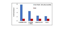
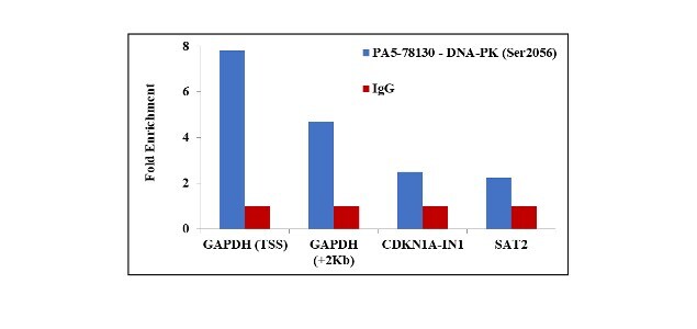
- Chromatin Immunoprecipitation (ChIP) assay of endogenous Phospho DNA-PK (Ser2056) protein using Anti-Phospho-DNA-PK (Ser2056) Antibody: ChIP was performed using Anti-Phospho-DNA-PK (Ser2056) Polyclonal Antibody (Product # PA5-78130, 2.5 µg) on sheared chromatin from camptothecin treated HeLa cells using the MAGnify ChIP System kit (Product # 49-2024). Normal Rabbit IgG was used as a negative IP control. The purified DNA was analyzed by qPCR using primers binding to GAPDH transcriptional start site, GAPDH gene body (+2Kb), CDKN1A intron 1 and SAT2 satellite repeats. Data is presented as fold enrichment of the antibody signal versus the negative control IgG using the comparative CT method.
Supportive validation
- Submitted by
- Invitrogen Antibodies (provider)
- Main image

- Experimental details
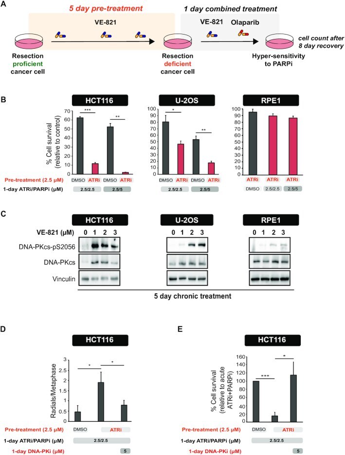
- Figure 4. Long-term ATR inhibition induces hypersensitivity to PARPi and hyperactivation of DNA-PKcs in cancer cell lines. ( A ) Experimental workflow of the assay used to measure cell survival after long-term ATRi. ( B ) HCT116, U-2OS and RPE1 cells were treated as shown in the schematic in ( A ). Cell viability was measured relative to cells treated with DMSO throughout all the pre-treatment and treatment period. Mean +- SD ( n = 3); * P < 0.05, ** P < 0.01, *** P < 0.001. ( C ) Levels of DNA-PKcs-pS2056 and total DNA-PKcs in cells after a 5-day treatment with the indicated concentrations of VE-821. ( D ) HCT116 cells were treated as in ( A ) for 5 days. After 5 days, cells were treated with olaparib (5 muM) with or without NU7441 (5 muM) for additional 24 hours. Metaphase spreads were then prepared as described in the 'Materials and Methods' section. Mean +- SD ( n = 3); * P < 0.05. ( E ) HCT116 cells were treated as in ( D ). Cell viability was measured relative to cells treated with acute ATRi and PARPi. Mean +- SD ( n = 4); * P < 0.05, *** P < 0.001.