 Explore
 Validate
 Learn
Learn Western blot
Antibody data
- Antibody Data
- Antigen structure
- References [1]
- Comments [0]
- Validations
- Western blot [2]
- Immunocytochemistry [1]
- Immunohistochemistry [1]
- Other assay [2]
Submit
Validation data
Reference
Comment
Report error
- Product number
- PA5-60685 - Provider product page

- Provider
- Invitrogen Antibodies
- Product name
- DHX40 Polyclonal Antibody
- Antibody type
- Polyclonal
- Antigen
- Recombinant full-length protein
- Description
- Immunogen sequence: SVGRTFCTMD GRGSPVHIHP SSALHEQETK LEWIIFHEVL VTTKVYARIV CPIRYEWVRD LLPKLHEFNA HDLSSVARRE VREDARRRWT NKENVKQLKD GISKDVLKKM QRRNDDKSIS DARARF
- Concentration
- 0.05 mg/mL
Submitted references Identification and Characterization of USP7 Targets in Cancer Cells.
Georges A, Marcon E, Greenblatt J, Frappier L
Scientific reports 2018 Oct 26;8(1):15833
Scientific reports 2018 Oct 26;8(1):15833
No comments: Submit comment
Supportive validation
- Submitted by
- Invitrogen Antibodies (provider)
- Main image

- Experimental details
- Western blot analysis of DHX40 in Lane 1: NIH-3T3 cell lysate (Mouse embryonic fibroblast cells); Lane 2: NBT-II cell lysate (Rat Wistar bladder tumour cells). Samples were probed using a DHX40 Polyclonal Antibody (Product # PA5-60685).

- Submitted by
- Invitrogen Antibodies (provider)
- Main image

- Experimental details
- Western blot analysis of DHX40 in Lane 1: Marker (kDa) 250, 130, 95, 72, 55, 36, 28, 17, 10; Lane 2: Human cell line RT-4; Lane 3: Human cell line U-251MG sp. Samples were probed using a DHX40 Polyclonal Antibody (Product # PA5-60685).

Supportive validation
- Submitted by
- Invitrogen Antibodies (provider)
- Main image

- Experimental details
- Immunofluorescent staining of DHX40 in human cell line U-251 MG shows positivity in nucleus but excluded from the nucleoli. Samples were probed using a DHX40 Polyclonal Antibody (Product # PA5-60685).

Supportive validation
- Submitted by
- Invitrogen Antibodies (provider)
- Main image

- Experimental details
- Immunohistochemical staining of DHX40 in human testis tissue shows strong nuclear and cytoplasmic positivity in Leydig cells and moderate nuclear staining in subsets of cells in seminiferus ducts. Samples were probed using a DHX40 Polyclonal Antibody (Product # PA5-60685).

Supportive validation
- Submitted by
- Invitrogen Antibodies (provider)
- Main image
- Experimental details
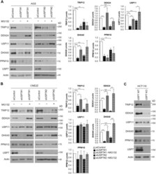
- Figure 4 Effects of USP7 depletion on target protein levels. ( A , B ) AGS ( A ) or CNE2Z ( B ) cells were transfected with two different siRNAs targeting USP7 (#1 or #2) or a negative control siRNA (siControl) followed by treatment with the MG132 proteasome inhibitor (+) or DMSO as a negative control (-). Cell lysates were analyzed by Western blotting using the indicated antibodies. For each condition, the protein bands for TRIP12, DDX24, USP11, DHX40 and PPM1G were quantified in three independent USP7 silencing experiments (+/-MG132) and normalized to actin. The bar graphs to the right of the Western blots show the average values relative to the silencing control for each protein. P values for siUSP7#1 or 2 are indicated relative to siControl and p values for siUSP7#1/2 + MG132 are indicated relative to siUSP7#1/2 without MG132 (*0.01 < P < 0.05; **0.001 < P < 0.01; *** P < 0.001). ( C ) Whole cell lysates of HCT116 cells (WT) or HCT116 with USP7 knockout (KO) were analyzed by Western blotting as in ( A ).

- Submitted by
- Invitrogen Antibodies (provider)
- Main image

- Experimental details
- Figure 1 Coimmunoprecipitation of USP7 target proteins with WT and binding pocket mutations of USP7. ( A ) AGS cell were transfected with a plasmid expressing myc-tagged USP7 or an empty myc plasmid control (VC). Myc-USP7 was immunoprecipitated with anti-myc resin and recovered proteins were analyzed by Western blotting using antibodies against myc and the indicated endogenous proteins. ( B ) CNE2Z cells were transfected with empty vector control (VC) plasmid or plasmids expressing myc-tagged USP7 with WT sequence or with mutations in the TRAF (DW), or Ubl2 binding pocket or both binding pockets (DW/Ubl2). Myc-USP7 was recovered by Myc immunoprecipitation, followed by Western blotting as in ( A ).
