 Explore
 Validate
 Learn
Learn Western blot
Antibody data
- Antibody Data
- Antigen structure
- References [5]
- Comments [0]
- Validations
- Western blot [1]
- Immunohistochemistry [3]
- Other assay [4]
Submit
Validation data
Reference
Comment
Report error
- Product number
- PA1-29421 - Provider product page

- Provider
- Invitrogen Antibodies
- Product name
- TRPV1 Polyclonal Antibody
- Antibody type
- Polyclonal
- Antigen
- Synthetic peptide
- Description
- Applications Reported: This CD28.2 antibody has been reported for use in flow cytometric analysis.
- Reactivity
- Human, Rat
- Host
- Rabbit
- Isotype
- IgG
- Vial size
- 50 µL
- Concentration
- Conc. Not Determined
- Storage
- Store at 4°C short term. For long term storage, store at -20°C, avoiding freeze/thaw cycles.
Submitted references Yellow Chaste Weed and Its Components, Apigenin and Galangin, Affect Proliferation and Oxidative Stress in Blue Light-Irradiated HaCaT Cells.
Dose-dependent immunomodulatory effects of bortezomib in experimental autoimmune neuritis.
Intrathecal injection of TRPV1 shRNA leads to increases in blood pressure in rats.
Enhanced salt sensitivity following shRNA silencing of neuronal TRPV1 in rat spinal cord.
Brain-derived neurotrophic factor redistribution in the dorsal root ganglia correlates with neuropathic pain inhibition after resiniferatoxin treatment.
Park JY, Park SH, Oh SW, Kwon K, Yu E, Choi S, Yang S, Han SB, Jung K, Song M, Cho JY, Lee J
Nutrients 2022 Mar 13;14(6)
Nutrients 2022 Mar 13;14(6)
Dose-dependent immunomodulatory effects of bortezomib in experimental autoimmune neuritis.
Klimas R, Sgodzai M, Motte J, Mohamad N, Renk P, Blusch A, Grüter T, Pedreiturria X, Gobrecht P, Fischer D, Schneider-Gold C, Reinacher-Schick A, Tannapfel A, Yoon MS, Gold R, Pitarokoili K
Brain communications 2021;3(4):fcab238
Brain communications 2021;3(4):fcab238
Intrathecal injection of TRPV1 shRNA leads to increases in blood pressure in rats.
Yu SQ, Wang DH
Acta physiologica (Oxford, England) 2011 Sep;203(1):139-47
Acta physiologica (Oxford, England) 2011 Sep;203(1):139-47
Enhanced salt sensitivity following shRNA silencing of neuronal TRPV1 in rat spinal cord.
Yu SQ, Wang DH
Acta pharmacologica Sinica 2011 Jun;32(6):845-52
Acta pharmacologica Sinica 2011 Jun;32(6):845-52
Brain-derived neurotrophic factor redistribution in the dorsal root ganglia correlates with neuropathic pain inhibition after resiniferatoxin treatment.
Tender GC, Li YY, Cui JG
The spine journal : official journal of the North American Spine Society 2010 Aug;10(8):715-20
The spine journal : official journal of the North American Spine Society 2010 Aug;10(8):715-20
No comments: Submit comment
Supportive validation
- Submitted by
- Invitrogen Antibodies (provider)
- Main image

- Experimental details
- HEK293 cells expressing combinations of TRPV1, p75, and trkA, as indicated in the figure, were treated with either NGF or vehicle and then immunoprecipitated with anti-trkA (lanes 1-7) or anti-TRPV1 (lanes 8-13) antibodies. Lanes 1-7 were separated from lanes 8-13 and the membranes were probed with anti-trkA antibody and TRPV1 Polyclonal Antibody (Product # PA1-29421), respectively (top). Segments of the membrane were then reapposed for imaging. The membranes were then stripped and reprobed with anti-phosphotyrosine antibody (bottom).

Supportive validation
- Submitted by
- Invitrogen Antibodies (provider)
- Main image

- Experimental details
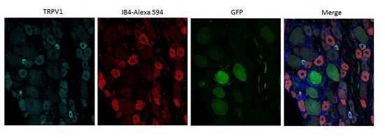
- Immunohistochemistry analysis of dorsal root ganglion using TRPV1 Polyclonal Antibody (Product # PA1-29421).

- Submitted by
- Invitrogen Antibodies (provider)
- Main image
- Experimental details
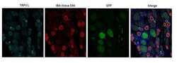
- Immunohistochemistry analysis of rat dorsal root ganglion using TRPV1 Polyclonal Antibody (Product # PA1-29421). Some DRG neurons express GFP (because they were infected by intrathecal administration of AAV2-5 carrying GFP). Cyan: Primary antibody. Red: IB4.
- Submitted by
- Invitrogen Antibodies (provider)
- Main image

- Experimental details
- Immunohistochemistry (Frozen) analysis of rat DRG tissue using (Product # PA1-29421) TRPV1 Polyclonal Antibody. Green: Primary antibody. Red: NeuN. Dilution: 1:1,000.

Supportive validation
- Submitted by
- Invitrogen Antibodies (provider)
- Main image

- Experimental details
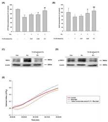
- Figure 3 Western blot analysis showing TRPV1 expression in the dorsal horn of the spinal cord (T8-L3), DRG (T8-L3), and mesenteric arteries of rats fed a NS or HS diet 3 d after control or TRPV1 shRNA treatment. The left, middle, and right panels indicate representative Western blots, quantification results (beta-actin arbitrary units), and the percentage change of TRPV1 expression of TRPV1 shRNA from that of corresponding control shRNA, respectively. Values are expressed as mean+-SEM ( n =5). b P

- Submitted by
- Invitrogen Antibodies (provider)
- Main image

- Experimental details
- NULL

- Submitted by
- Invitrogen Antibodies (provider)
- Main image
- Experimental details
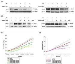
- Figure 1 YCW extract increased cell proliferation via calcium dependent-TRPV1 signaling pathways in blue light irradiated HaCaT cells. ( A ) CellTiter-Glo (r) 2.0 assay of YCW extract (0.01-0.1%) pretreated with blue light-irradiated HaCaT cells. YCW extract was pretreated 24 h before irradiation of blue light (13.68 J/cm 2 ), and the same process was repeated on the next day. After 24 h incubation, an equal volume of CellTiter-Glo reagent was added to the cells. ( B ) Cell proliferation effect of YCW extract (0.01-0.1%) on blue light-irradiated HaCaT cells was measured using BrdU ELISA assay. Data are presented as the mean +- SEM of four independent experiments. Statistical significance of differences among the groups was assessed by one-way analysis of variance (ANOVA), followed by Tukey's multiple-comparison test, using the GraphPad Prism 5 software. * p < 0.05 vs. control group. * p < 0.05 vs. control, *** p < 0.005 vs. control, # p < 0.05 vs. blue light-irradiated group (B.L), ## p < 0.01 vs. B.L. ### p < 0.005 vs. B.L. ( C ) TRPV1 expression levels were determined by Western blotting at 24 h incubation after, pretreatment with YCW extract (0.1%) for and 2-day repetitive blue light irradiation (30 min, 76 W/m 2 ). ( D ) Phosphorylated-TRPV1 expression levels were determined by Western blotting at 90 min incubation after, 1 h YCW extract (0.1%) pretreatment and blue light irradiation (30 min, 76 W/m 2 ). The total proteins were extracted from the cells immediately after e

- Submitted by
- Invitrogen Antibodies (provider)
- Main image
- Experimental details
- Apigenin and galangin reduced TRPV1 expression and its phosphorylation in blue light-irradiated HaCaT cells. ( A , B ) TRPV1 expression ( A ), and its phosphorylation levels ( B ) were determined by Western blotting. ( A ) Cells were incubated with apigenin (10, 20, 30 muM) and galangin (1, 10, 20 muM) for 24 h and were then irradiated with blue light (30 min, 76 W/m 2 ). After 24 h incubation, the cells were subjected to the same process twice and were finally subjected to Western blot analysis. ( B ) Phosphorylation levels of TRPV1 were determined by Western blotting at 30-, 60-, and 90-min incubation after pretreatment with apigenin (30 muM) or galangin (20 muM) for 1 h and subsequent blue light-irradiation (30 min, 76 W/m 2 ). The total proteins were extracted from the cells immediately after experimental conditions, and beta-actin was used as a loading control. ( C , D ) Apigenin and galangin inhibited blue light-induced calcium influx in HaCaT cells. ( C ) Ca 2+ influx changes of blue light and with apigenin (10, 20, 30 muM) pretreatment, ( D ) with galangin (1, 10, 20 muM) pretreatment by Fluo-4 NW assay. Cells were pretreated with apigenin (10, 20, 30 muM) and galangin (1, 10, 20 muM) for 1 h and irradiated with blue light (10 min, 76 W/m 2 ). After the irradiation, fluorescence intensities were measured immediately over a certain period.
