 Explore
 Validate
 Learn
Learn Western blot
Antibody data
- Antibody Data
- Antigen structure
- References [0]
- Comments [0]
- Validations
- Western blot [2]
- Immunocytochemistry [1]
- Immunohistochemistry [1]
Submit
Validation data
Reference
Comment
Report error
- Product number
- TA328624 - Provider product page

- Provider
- OriGene
- Product name
- Rabbit Polyclonal Anti-Melanocortin Receptor 1
- Antibody type
- Polyclonal
- Description
- Rabbit Polyclonal Anti-Melanocortin Receptor 1
- Host
- Rabbit
- Conjugate
- Unconjugated
- Epitope
- MC1R
- Antibody clone number
- NULL
- Vial size
- 200 µl
- Concentration
- NULL
No comments: Submit comment
Supportive validation
- Submitted by
- OriGene (provider)
- Main image
- Experimental details
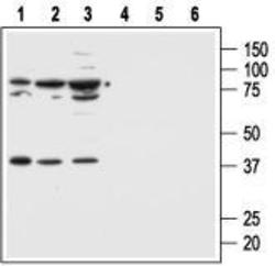
- Western blot analysis of human normal skin fibroblast cell line Malme-3 (lanes 1 and 4) and human malignant melanoma cell lines Malme-3M (lanes 2 and 5) and A875 (lanes 3 and 6): 1, 2, 3. Anti-Melanocortin Receptor 1 antibody, (1:500). 4, 5, 6. Anti-Melanocortin Receptor 1 antibody, preincubated with the control peptide antigen
- Validation comment
- WB

- Submitted by
- OriGene (provider)
- Main image
- Experimental details
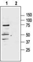
- Western blot analysis of rat adrenal lysate: 1. Anti-Melanocortin Receptor 1 antibody, (1:400). 2. Anti-Melanocortin Receptor 1 antibody, preincubated with the control peptide antigen.
- Validation comment
- WB

Supportive validation
- Submitted by
- OriGene (provider)
- Main image
- Experimental details
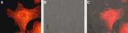
- Expression of MC1R in human Malme-3M cells. Immunocytochemical staining of human paraformaldehyde fixed and permeabilized malignant melanoma cell lines (Malme-3M). A. Cells were stained with Anti-Melanocortin Receptor 1 antibody, (1:200) followed by goat-anti-rabbit- AlexaFluor-555 secondary antibody. B. Live view of the same field as in (A). C. Computer merge of (A) and (B).
- Validation comment
- IF

Supportive validation
- Submitted by
- OriGene (provider)
- Main image

- Experimental details
- Expression of MC1R in normal skin and melanoma. Immunohistochemical staining of paraffin embedded normal skin and melanoma sections using Anti-Melanocortin Receptor 1 antibody(1:100). MCR1 staining (red-brown color is highly specific in A. epidermal cells, B. eccrine sweat gland cells and C. melanoma cells. Color reaction was obtained with DAB. Hematoxilin is used as the counterstain.
- Validation comment
- IHC
