Explore
 Validate
 Learn
Learn Western blot
Antibody data
- Antibody Data
- Antigen structure
- References [15]
- Comments [0]
- Validations
- Western blot [1]
- Immunocytochemistry [1]
- Flow cytometry [1]
Submit
Validation data
Reference
Comment
Report error
- Product number
- AF3688 - Provider product page

- Provider
- R&D Systems
- Product name
- Mouse LRIG1 Antibody
- Antibody type
- Polyclonal
- Description
- Immunogen affinity purified. Detects mouse LRIG1 in direct ELISAs and Western blots. In direct ELISAs, less than 10% cross-reactivity with recombinant human (rh) LRIG1 is observed and less than 5% cross-reactivity with rhLRIG3 is observed.
- Reactivity
- Mouse
- Host
- Goat
- Conjugate
- Unconjugated
- Antigen sequence
P70193- Isotype
- IgG
- Vial size
- 100 ug
- Concentration
- LYOPH
- Storage
- Use a manual defrost freezer and avoid repeated freeze-thaw cycles. 12 months from date of receipt, -20 to -70 °C as supplied. 1 month, 2 to 8 °C under sterile conditions after reconstitution. 6 months, -20 to -70 °C under sterile conditions after reconstitution.
Submitted references YAP/TAZ-Dependent Reprogramming of Colonic Epithelium Links ECM Remodeling to Tissue Regeneration.
JunB defines functional and structural integrity of the epidermo-pilosebaceous unit in the skin.
Secretory phospholipase A2-IIA overexpressing mice exhibit cyclic alopecia mediated through aberrant hair shaft differentiation and impaired wound healing response.
Stem Cell Lineage Infidelity Drives Wound Repair and Cancer.
Stem cell plasticity enables hair regeneration following Lgr5+ cell loss.
GPR39 marks specific cells within the sebaceous gland and contributes to skin wound healing.
Overexpression of epigen during embryonic development induces reversible, epidermal growth factor receptor-dependent sebaceous gland hyperplasia.
Kindlin-1 controls Wnt and TGF-β availability to regulate cutaneous stem cell proliferation.
Distinct fibroblast lineages determine dermal architecture in skin development and repair.
Transcription factor Oct1 is a somatic and cancer stem cell determinant.
Delayed cutaneous wound healing and aberrant expression of hair follicle stem cell markers in mice selectively lacking Ctip2 in epidermis.
Lrig1 controls intestinal stem-cell homeostasis by negative regulation of ErbB signalling.
TCF/Lef1 activity controls establishment of diverse stem and progenitor cell compartments in mouse epidermis.
Assaying proliferation and differentiation capacity of stem cells using disaggregated adult mouse epidermis.
Differential sensitivity of epidermal cell subpopulations to beta-catenin-induced ectopic hair follicle formation.
Yui S, Azzolin L, Maimets M, Pedersen MT, Fordham RP, Hansen SL, Larsen HL, Guiu J, Alves MRP, Rundsten CF, Johansen JV, Li Y, Madsen CD, Nakamura T, Watanabe M, Nielsen OH, Schweiger PJ, Piccolo S, Jensen KB
Cell stem cell 2018 Jan 4;22(1):35-49.e7
Cell stem cell 2018 Jan 4;22(1):35-49.e7
JunB defines functional and structural integrity of the epidermo-pilosebaceous unit in the skin.
Singh K, Camera E, Krug L, Basu A, Pandey RK, Munir S, Wlaschek M, Kochanek S, Schorpp-Kistner M, Picardo M, Angel P, Niemann C, Maity P, Scharffetter-Kochanek K
Nature communications 2018 Aug 24;9(1):3425
Nature communications 2018 Aug 24;9(1):3425
Secretory phospholipase A2-IIA overexpressing mice exhibit cyclic alopecia mediated through aberrant hair shaft differentiation and impaired wound healing response.
Chovatiya GL, Sarate RM, Sunkara RR, Gawas NP, Kala V, Waghmare SK
Scientific reports 2017 Sep 14;7(1):11619
Scientific reports 2017 Sep 14;7(1):11619
Stem Cell Lineage Infidelity Drives Wound Repair and Cancer.
Ge Y, Gomez NC, Adam RC, Nikolova M, Yang H, Verma A, Lu CP, Polak L, Yuan S, Elemento O, Fuchs E
Cell 2017 May 4;169(4):636-650.e14
Cell 2017 May 4;169(4):636-650.e14
Stem cell plasticity enables hair regeneration following Lgr5+ cell loss.
Hoeck JD, Biehs B, Kurtova AV, Kljavin NM, de Sousa E Melo F, Alicke B, Koeppen H, Modrusan Z, Piskol R, de Sauvage FJ
Nature cell biology 2017 Jun;19(6):666-676
Nature cell biology 2017 Jun;19(6):666-676
GPR39 marks specific cells within the sebaceous gland and contributes to skin wound healing.
Zhao H, Qiao J, Zhang S, Zhang H, Lei X, Wang X, Deng Z, Ning L, Cao Y, Guo Y, Liu S, Duan E
Scientific reports 2015 Jan 21;5:7913
Scientific reports 2015 Jan 21;5:7913
Overexpression of epigen during embryonic development induces reversible, epidermal growth factor receptor-dependent sebaceous gland hyperplasia.
Dahlhoff M, Frances D, Kloepper JE, Paus R, Schäfer M, Niemann C, Schneider MR
Molecular and cellular biology 2014 Aug;34(16):3086-95
Molecular and cellular biology 2014 Aug;34(16):3086-95
Kindlin-1 controls Wnt and TGF-β availability to regulate cutaneous stem cell proliferation.
Rognoni E, Widmaier M, Jakobson M, Ruppert R, Ussar S, Katsougkri D, Böttcher RT, Lai-Cheong JE, Rifkin DB, McGrath JA, Fässler R
Nature medicine 2014 Apr;20(4):350-9
Nature medicine 2014 Apr;20(4):350-9
Distinct fibroblast lineages determine dermal architecture in skin development and repair.
Driskell RR, Lichtenberger BM, Hoste E, Kretzschmar K, Simons BD, Charalambous M, Ferron SR, Herault Y, Pavlovic G, Ferguson-Smith AC, Watt FM
Nature 2013 Dec 12;504(7479):277-281
Nature 2013 Dec 12;504(7479):277-281
Transcription factor Oct1 is a somatic and cancer stem cell determinant.
Maddox J, Shakya A, South S, Shelton D, Andersen JN, Chidester S, Kang J, Gligorich KM, Jones DA, Spangrude GJ, Welm BE, Tantin D
PLoS genetics 2012;8(11):e1003048
PLoS genetics 2012;8(11):e1003048
Delayed cutaneous wound healing and aberrant expression of hair follicle stem cell markers in mice selectively lacking Ctip2 in epidermis.
Liang X, Bhattacharya S, Bajaj G, Guha G, Wang Z, Jang HS, Leid M, Indra AK, Ganguli-Indra G
PloS one 2012;7(2):e29999
PloS one 2012;7(2):e29999
Lrig1 controls intestinal stem-cell homeostasis by negative regulation of ErbB signalling.
Wong VW, Stange DE, Page ME, Buczacki S, Wabik A, Itami S, van de Wetering M, Poulsom R, Wright NA, Trotter MW, Watt FM, Winton DJ, Clevers H, Jensen KB
Nature cell biology 2012 Mar 4;14(4):401-8
Nature cell biology 2012 Mar 4;14(4):401-8
TCF/Lef1 activity controls establishment of diverse stem and progenitor cell compartments in mouse epidermis.
Petersson M, Brylka H, Kraus A, John S, Rappl G, Schettina P, Niemann C
The EMBO journal 2011 Jun 21;30(15):3004-18
The EMBO journal 2011 Jun 21;30(15):3004-18
Assaying proliferation and differentiation capacity of stem cells using disaggregated adult mouse epidermis.
Jensen KB, Driskell RR, Watt FM
Nature protocols 2010 May;5(5):898-911
Nature protocols 2010 May;5(5):898-911
Differential sensitivity of epidermal cell subpopulations to beta-catenin-induced ectopic hair follicle formation.
Baker CM, Verstuyf A, Jensen KB, Watt FM
Developmental biology 2010 Jul 1;343(1-2):40-50
Developmental biology 2010 Jul 1;343(1-2):40-50
No comments: Submit comment
Supportive validation
- Submitted by
- R&D Systems (provider)
- Main image

- Experimental details
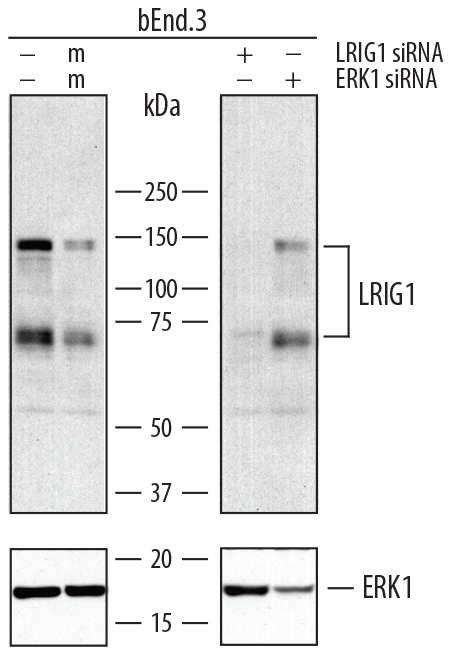
- Detection of Mouse LRIG1 by Western Blot. Western blot shows lysates of bEnd.3 mouse endothelioma cell line untreated (-), treated (+) or mock treated (m) with 30 pmol mouse LRIG1 or mouse ERK1 siRNA. PVDF membrane was probed with 2 µg/mL of Goat Anti-Mouse LRIG1 Antigen Affinity-purified Polyclonal Antibody (Catalog # AF3688) followed by HRP-conjugated Anti-Goat IgG Secondary Antibody (Catalog # HAF109). Specific bands were detected for LRIG1 at approximately 140 kDa and 70 kDa (as indicated, upper panel). For additional reference ERK1 was detected using Rabbit Anti-Human/Mouse/Rat ERK1 Antigen Affinity-purified Polyclonal Antibody (lower panel, Catalog # AF1575) This experiment was conducted under reducing conditions and using Immunoblot Buffer Group 1.
Supportive validation
- Submitted by
- R&D Systems (provider)
- Main image

- Experimental details
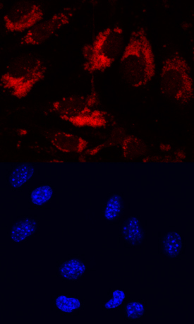
- LRIG1 in bEnd.3 Mouse Cell Line. LRIG1 was detected in immersion fixed bEnd.3 mouse endothelioma cell line using Goat Anti-Mouse LRIG1 Antigen Affinity-purified Polyclonal Antibody (Catalog # AF3688) at 10 µg/mL for 3 hours at room temperature. Cells were stained using the NorthernLights™ 557-conjugated Anti-Goat IgG Secondary Antibody (red, upper panel; Catalog # NL001) and counterstained with DAPI (blue, lower panel). Specific staining was localized to cytoplasm. View our protocol for Fluorescent ICC Staining of Cells on Coverslips.
Supportive validation
- Submitted by
- R&D Systems (provider)
- Main image

- Experimental details
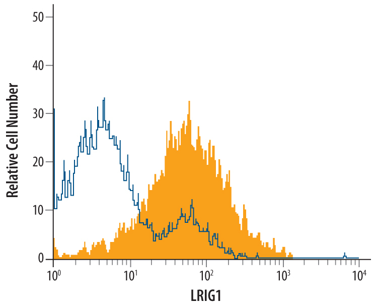
- Detection of LRIG1 in bEnd.3 Mouse Cell Line by Flow Cytometry. bEnd.3 mouse endothelioma cell line was stained with Goat Anti-Mouse LRIG1 Antigen Affinity-purified Polyclonal Antibody (Catalog # AF3688, filled histogram) or control antibody (Catalog # AB-108-C, open histogram), followed by Phycoerythrin-conjugated Anti-Goat IgG Secondary Antibody (Catalog # F0107).