Antibody data
- Antibody Data
- Antigen structure
- References [1]
- Comments [0]
- Validations
- Western blot [2]
- Immunohistochemistry [2]
- Other assay [1]
Submit
Validation data
Reference
Comment
Report error
- Product number
- PA5-36552 - Provider product page

- Provider
- Invitrogen Antibodies
- Product name
- Aquaporin 3 Polyclonal Antibody
- Antibody type
- Polyclonal
- Antigen
- Synthetic peptide
- Description
- This antibody detects endogenous protein at a molecular weight of 32 kDa. Purity is >95% by SDS-PAGE.
- Reactivity
- Human, Mouse, Rat
- Host
- Rabbit
- Isotype
- IgG
- Vial size
- 100 µL
- Concentration
- 1 mg/mL
- Storage
- Store at 4°C short term. For long term storage, store at -20°C, avoiding freeze/thaw cycles.
Submitted references Mitochondrial dysfunction promotes aquaporin expression that controls hydrogen peroxide permeability and ferroptosis.
Takashi Y, Tomita K, Kuwahara Y, Roudkenar MH, Roushandeh AM, Igarashi K, Nagasawa T, Nishitani Y, Sato T
Free radical biology & medicine 2020 Dec;161:60-70
Free radical biology & medicine 2020 Dec;161:60-70
No comments: Submit comment
Supportive validation
- Submitted by
- Invitrogen Antibodies (provider)
- Main image

- Experimental details
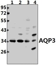
- Western blot analysis of Aquaporin 3 in Lane 1: HEK293T whole cell lysate (40 µg), Lane 2: A375 whole cell lysate (40 µg), Lane 3: the Kidney tissue lysate of mouse (40 µg), Lane 4: the Kidney tissue lysate of rat (40 µg). Samples were incubated with Aquaporin 3 polyclonal antibody (Product # PA5-36552) at a dilution of 1:500.
- Submitted by
- Invitrogen Antibodies (provider)
- Main image

- Experimental details
- Western blot analysis of Aquaporin-3 using Aquaporin-3 polyclonal antibody (Product # PA5-36552) at a dilution of 1:500. Lane 1: Hela cell lysate, Lane 2: HEK293T cell lysate, Lane 3: NIH-3T3 cell lysate.
Supportive validation
- Submitted by
- Invitrogen Antibodies (provider)
- Main image

- Experimental details
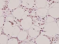
- Immunohistochemistry analysis of Aquaporin 3 in paraffin-embedded human breast carcinoma tissue. Samples were incubated with Aquaporin 3 polyclonal antibody (Product # PA5-36552) at a dilution of 1:100.
- Submitted by
- Invitrogen Antibodies (provider)
- Main image

- Experimental details
- Immunohistochemical analysis of Aquaporin-3 in paraffin-embedded human breast carcinoma using Aquaporin-3 polyclonal antibody (Product # PA5-36552) at a dilution of 1:100.
Supportive validation
- Submitted by
- Invitrogen Antibodies (provider)
- Main image

- Experimental details
- Fig. 3 Spatial distribution of AQPs that function as H 2 O 2 channels. Immunostaining of AQPs was performed to investigate the contribution of AQPs to H 2 O 2 permeability. A: Immunostaining of AQP3 in HeLa and SAS rho 0 cells. B: Relative fluorescence intensity of AQP3 in HeLa and SAS rho 0 cells. C: Immunostaining of AQP5. D: Relative intensity of AQP5. E: Immunostaining of AQP8. F: Relative fluorescence intensity of AQP8. In HeLa and SAS rho 0 cells, AQPs were strongly expressed in the plasma membrane, and average expression intensities were significantly higher than in parental cells. **: p < 0.01 using Student's t- test (vs. parent). Fig. 3
 Explore
 Validate
 Learn
Learn Western blot