Explore
 Validate
 Learn
Learn Western blot
Antibody data
- Antibody Data
- Antigen structure
- References [0]
- Comments [0]
- Validations
- Western blot [4]
- Immunohistochemistry [1]
- Flow cytometry [1]
Submit
Validation data
Reference
Comment
Report error
- Product number
- NBP2-29942 - Provider product page

- Provider
- Novus Biologicals
- Product name
- Rabbit Polyclonal CYP27B1 Antibody
- Antibody type
- Polyclonal
- Description
- Ammonium sulfate precipitation.
- Reactivity
- Human, Rat
- Host
- Rabbit
- Isotype
- IgG
- Vial size
- 0.4 ml
- Concentration
- 6.9 mg/ml
- Storage
- Store at 4C short term. Aliquot and store at -20C long term. Avoid freeze-thaw cycles.
No comments: Submit comment
Supportive validation
- Submitted by
- Novus Biologicals (provider)
- Main image

- Experimental details
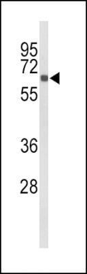
- Western Blot: CYP27B1 Antibody [NBP2-29942] - Western blot analysis of (C-term) (NBP2-29942) in mouse kidney tissue lysates (35ug/lane). CYP27B1 (arrow) was detected using the purified Pab.
- Submitted by
- Novus Biologicals (provider)
- Main image
- Experimental details
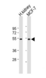
- Western Blot: CYP27B1 Antibody [NBP2-29942] - Antibody (C-term) at 1:2000 dilution Lane 1: Human kidney lysate Lane 2: MCF-7 whole cell lysate Lysates/proteins at 20 ug per lane. Secondary Goat Anti-Rabbit IgG, (H+L), Peroxidase conjugated at 1/10000 dilution. Predicted band size : 57 kDa Blocking/Dilution buffer: 5% NFDM/TBST.

- Submitted by
- Novus Biologicals (provider)
- Main image
- Experimental details
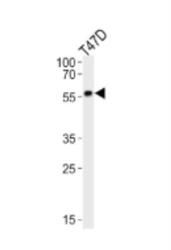
- Western Blot: CYP27B1 Antibody [NBP2-29942] - Analysis of lysate from T47D cell line, using CYP27B1 Antibody (C-term). Diluted at 1:1000. A goat anti-rabbit IgG H&L(HRP) at 1:10000 dilution was used as the secondary antibody. Lysate at 20ug.

- Submitted by
- Novus Biologicals (provider)
- Main image
- Experimental details
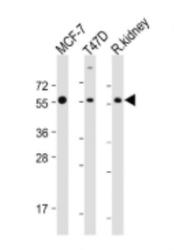
- Western Blot: CYP27B1 Antibody [NBP2-29942] - Antibody (C-term) at 1:2000 dilution Lane 1: MCF-7 whole cell lysate Lane 2: T47D whole cell lysate Lane 3: Rat kidney lysate Lysates/proteins at 20 ug per lane. Secondary Goat Anti-Rabbit IgG, (H+L), Peroxidase conjugated at 1/10000 dilution. Predicted band size : 57 kDa Blocking/Dilution buffer: 5% NFDM/TBST.

Supportive validation
- Submitted by
- Novus Biologicals (provider)
- Main image
- Experimental details
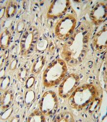
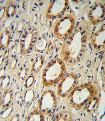
- Immunohistochemistry-Paraffin: CYP27B1 Antibody [NBP2-29942] - Immunohistochemistry analysis in formalin fixed and paraffin embedded human kidney tissue followed by peroxidase conjugation of the secondary antibody and DAB staining.This data demonstrates the use of (C-term) for immunohistochemistry. Clinical relevance has not been evaluated.
Supportive validation
- Submitted by
- Novus Biologicals (provider)
- Main image
- Experimental details
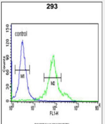
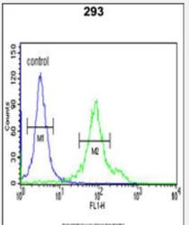
- Flow Cytometry: CYP27B1 Antibody [NBP2-29942] - Flow cytometric analysis of 293 cells (right histogram) compared to a negative control cell (left histogram).FITC-conjugated goat-anti-rabbit secondary antibodies were used for the analysis.