Explore
 Validate
 Learn
Learn Western blot
Antibody data
- Antibody Data
- Antigen structure
- References [2]
- Comments [0]
- Validations
- Western blot [2]
- Immunocytochemistry [2]
- Immunohistochemistry [2]
- Other assay [2]
Submit
Validation data
Reference
Comment
Report error
- Product number
- PA5-104508 - Provider product page

- Provider
- Invitrogen Antibodies
- Product name
- Collagen IV Polyclonal Antibody
- Antibody type
- Polyclonal
- Antigen
- Synthetic peptide
- Reactivity
- Human, Mouse, Rat
- Host
- Rabbit
- Isotype
- IgG
- Vial size
- 100 µL
- Concentration
- 1 mg/mL
- Storage
- -20°C
Submitted references Mouse models for dominant dystrophic epidermolysis bullosa carrying common human point mutations recapitulate the human disease.
Restoration of renal TIMP3 levels via genetics and pharmacological approach prevents experimental diabetic nephropathy.
Smith BRC, Nyström A, Nowell CJ, Hausser I, Gretzmeier C, Robertson SJ, Varigos GA, Has C, Kern JS, Pang KC
Disease models & mechanisms 2021 Jun 1;14(6)
Disease models & mechanisms 2021 Jun 1;14(6)
Restoration of renal TIMP3 levels via genetics and pharmacological approach prevents experimental diabetic nephropathy.
Casagrande V, Iuliani G, Menini S, Pugliese G, Federici M, Menghini R
Clinical and translational medicine 2021 Feb;11(2):e305
Clinical and translational medicine 2021 Feb;11(2):e305
No comments: Submit comment
Supportive validation
- Submitted by
- Invitrogen Antibodies (provider)
- Main image

- Experimental details
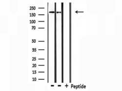
- Western blot analysis of Collagen IV in 293 and HeLa (Lane 1: 293 cells, Lane 2: Hela cells, Lane 3: 293 cells treated with blocking peptide). Samples were incubated with Collagen IV polyclonal antibody (Product # PA5-104508).

- Submitted by
- Invitrogen Antibodies (provider)
- Main image
- Experimental details
- Western blot analysis of Collagen IV in 293 and HeLa (Lane 1: 293 cells, Lane 2: Hela cells, Lane 3: 293 cells treated with blocking peptide). Samples were incubated with Collagen IV polyclonal antibody (Product # PA5-104508).

Supportive validation
- Submitted by
- Invitrogen Antibodies (provider)
- Main image
- Experimental details
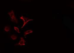
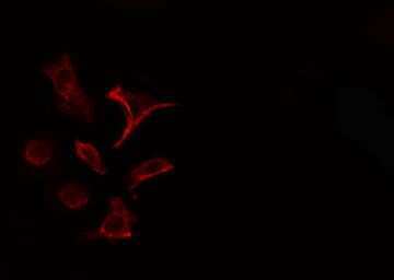
- Immunofluorescent analysis of Collagen IV in HeLa cells. Samples were fixed with paraformaldehyde, permeabilized with 0.1% Triton X-100, blocked with 10% serum (45 min at 25°C) incubated with Collagen IV polyclonal antibody (Product # PA5-104508) using a dilution of 1:200 (1 hr, 37°C), and followed by goat anti-rabbit IgG Alexa Fluor 594 at a dilution of 1:600.

- Submitted by
- Invitrogen Antibodies (provider)
- Main image

- Experimental details
- Immunofluorescent analysis of Collagen IV in HeLa cells. Samples were fixed with paraformaldehyde, permeabilized with 0.1% Triton X-100, blocked with 10% serum (45 min at 25°C) incubated with Collagen IV polyclonal antibody (Product # PA5-104508) using a dilution of 1:200 (1 hr, 37°C), and followed by goat anti-rabbit IgG Alexa Fluor 594 at a dilution of 1:600.
Supportive validation
- Submitted by
- Invitrogen Antibodies (provider)
- Main image

- Experimental details
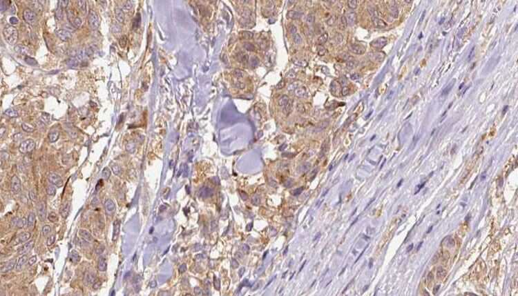
- Immunohistochemistry analysis of paraffin-embedded Collagen IV in human colon cancer tissues sections. Antigen retrieval was performed using citrate buffer. Samples were blocked with blocking buffer (1.5 hr, 22°C), incubated with Collagen IV polyclonal antibody (Product # PA5-104508) using a dilution of 1:100 (1.5 hr, 22°C), followed by HRP conjugated goat anti-rabbit.

- Submitted by
- Invitrogen Antibodies (provider)
- Main image

- Experimental details
- Immunohistochemistry analysis of paraffin-embedded Collagen IV in human Melanoma tissue. Antigen retrieval was performed using citrate buffer. Samples were blocked with blocking buffer (1.5 hr, 22°C), incubated with Collagen IV polyclonal antibody (Product # PA5-104508) using a dilution of 1:100 (1.5 hr, 22°C), followed by HRP conjugated goat anti-rabbit.
Supportive validation
- Submitted by
- Invitrogen Antibodies (provider)
- Main image

- Experimental details
- FIGURE 2 TIMP3 overexpression in macrophages reduces STZ-induced inflammation and fibrosis in kidney. Kidney cortex analysis of (A) gene expression of F4/80, MCP1, TNF-alpha, IL6, and COL1A2 and (B) protein levels of WT1, Podocin, Nephrin, Coll-IV, TGF-beta, Actin, phospho-p38 (p-p38), p38 in wt, and MacT3 non-diabetic and diabetic mice (n = 6 per group). (C) Protein levels of WT1, Podocin, and Actin in kidney glomerular fraction of wt and MacT3 non-diabetic and diabetic mice (n = 5 per group). A representative image of two mice per group is shown. Relative protein levels as determined by densitometry (*p < 0.05, **p

- Submitted by
- Invitrogen Antibodies (provider)
- Main image

- Experimental details
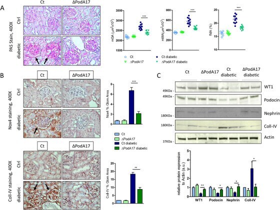
- FIGURE 4 Podocyte deletion of ADAM17 attenuates STZ-induced injury in kidney. Twelve weeks after STZ administration diabetic Ct and DeltaPodA17 mice and age-matched non-diabetic littermates (Ctrl) were analyzed for (A) PAS-staining in kidney and quantification of mGA, fMA, and mMA (arrows indicate glomerular hypertrophy and mesangial expansion) and (B) immunohistochemical staining of Nox4 and Coll-IV in kidney (non-diabetic n = 5, diabetic n = 10 per group, original magnification, X400) (arrows indicate increased glomerular staining for Nox4 and Coll-IV). (C) Protein expression of WT1, Podocin, Nephrin, Coll-IV, and Actin in kidney cortex from Ct and DeltaPodA17 non-diabetic and diabetic mice (n = 6 per group). A representative image of two mice per group is shown. Relative protein levels as determined by densitometry (*p < 0.05, **p