Explore
 Validate
 Learn
Learn Western blot
Antibody data
- Antibody Data
- Antigen structure
- References [1]
- Comments [0]
- Validations
- Western blot [2]
- Immunohistochemistry [2]
- Other assay [1]
Submit
Validation data
Reference
Comment
Report error
- Product number
- PA5-21058 - Provider product page

- Provider
- Invitrogen Antibodies
- Product name
- SHISA9 Polyclonal Antibody
- Antibody type
- Polyclonal
- Antigen
- Synthetic peptide
- Description
- A suggested positive control is rat brain tissue lysate. PA5-21058 can be used with blocking peptide PEP-1172.
- Reactivity
- Human, Mouse, Rat
- Host
- Rabbit
- Isotype
- IgG
- Vial size
- 100 µg
- Concentration
- 1 mg/mL
- Storage
- Maintain refrigerated at 2-8°C for up to 3 months. For long term storage store at -20°C
Submitted references C-terminal interactors of the AMPA receptor auxiliary subunit Shisa9.
Karataeva AR, Klaassen RV, Ströder J, Ruiperez-Alonso M, Hjorth JJ, van Nierop P, Spijker S, Mansvelder HD, Smit AB
PloS one 2014;9(2):e87360
PloS one 2014;9(2):e87360
No comments: Submit comment
Supportive validation
- Submitted by
- Invitrogen Antibodies (provider)
- Main image

- Experimental details
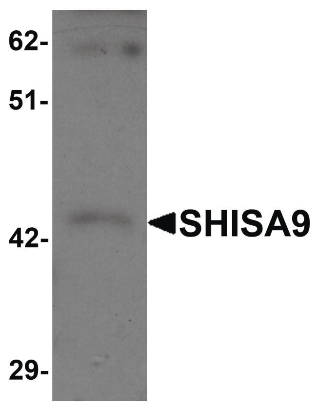
- Western blot analysis of rat brain tissue lysate using a SHISA9 polyclonal antibody (Product # PA5-21058) at 1 µg/mL.
- Submitted by
- Invitrogen Antibodies (provider)
- Main image

- Experimental details
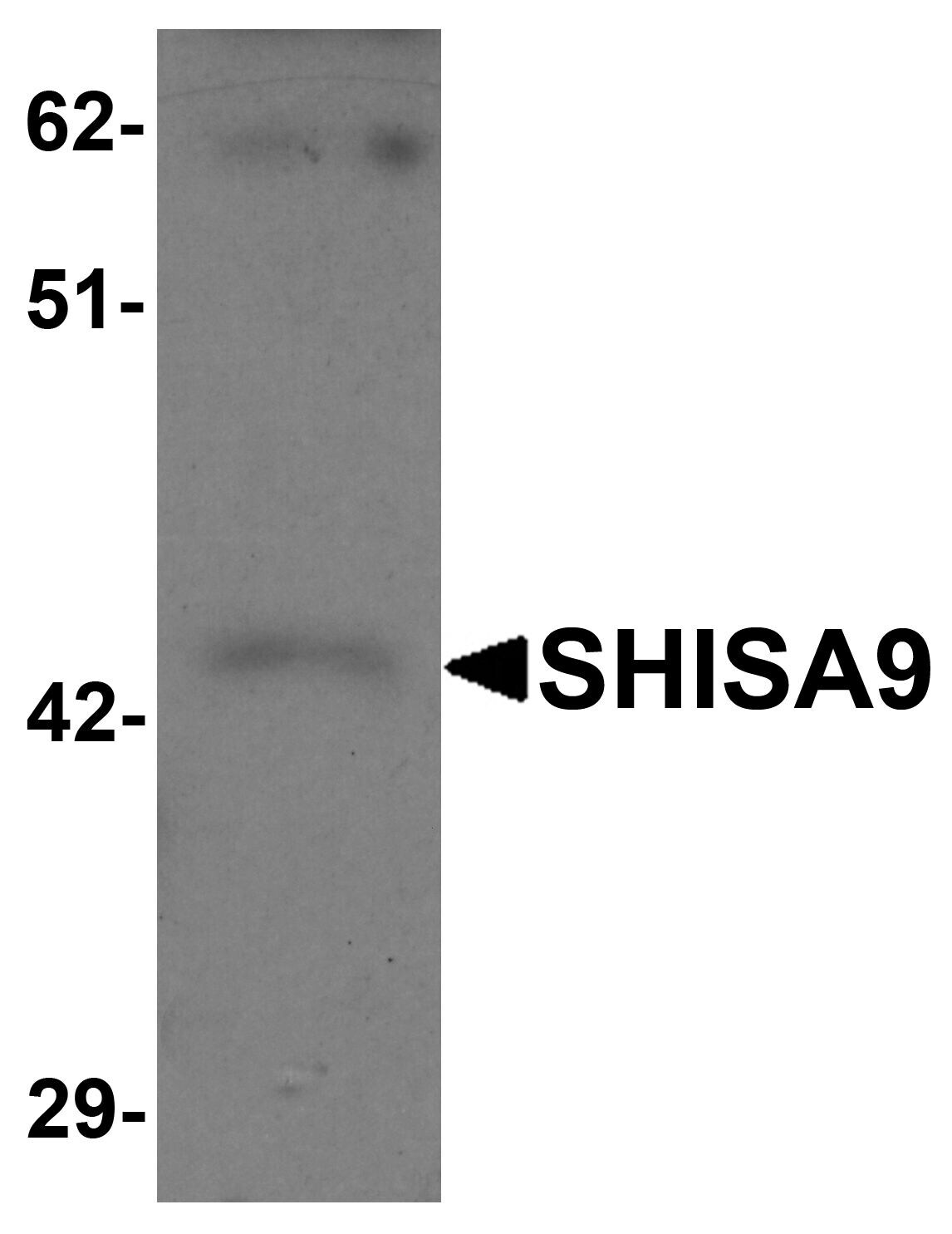
- Western Blot analysis of SHISA9 in rat brain tissue lysate with SHISA9 Polyclonal Antibody (Product # PA5-21058) at 1 µg/mL.
Supportive validation
- Submitted by
- Invitrogen Antibodies (provider)
- Main image

- Experimental details
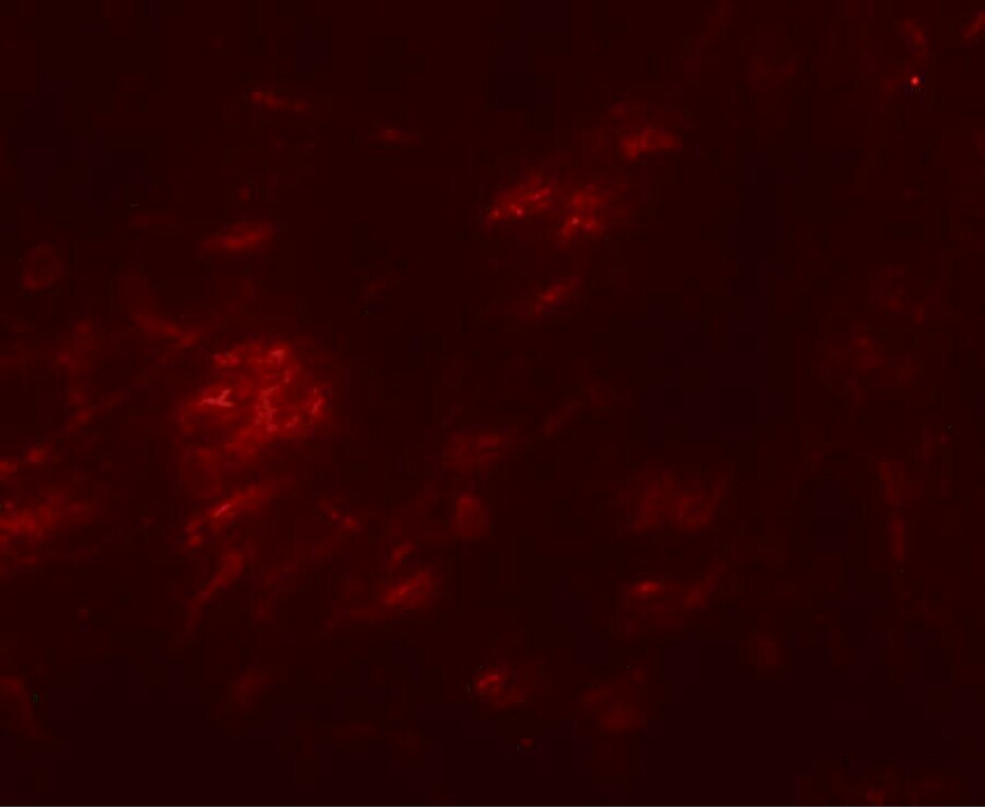
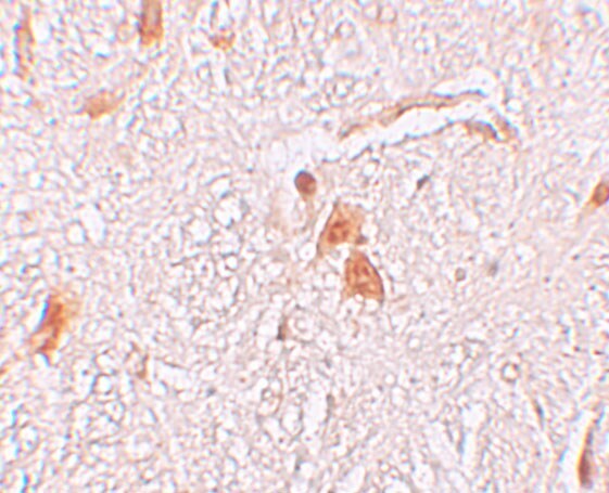
- Immunofluorescence of SHISA9 in human brain tissue with SHISA9 Polyclonal Antibody (Product # PA5-21058) at 20 µg/mL.
- Submitted by
- Invitrogen Antibodies (provider)
- Main image
- Experimental details
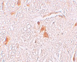
- Immunohistochemistry of SHISA9 in human brain tissue with SHISA9 Polyclonal Antibody (Product # PA5-21058) at 2.5 µg/mL.
Supportive validation
- Submitted by
- Invitrogen Antibodies (provider)
- Main image
- Experimental details
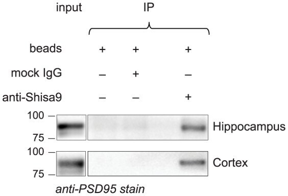
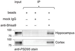
- Figure 3 Validation of the interaction of Shisa9-PSD95 the brain. Shisa9 forms a complex with PSD95 in the hippocampus and cortex. Anti-Shisa9 antibody was added to the mouse cortex and hippocampus lysates to immunoprecipitate native Shisa9 complexes. Obtained samples were resolved on SDS-PAGE, western blotted and immunostained with anti-PSD95 antibody. The 75 and 100 kDa bands are indicated.