 Explore
 Validate
 Learn
LearnHPA006431
antibody from Atlas Antibodies
Targeting: UBQLN2
Chap1, CHAP1/DSK2, Dsk2, LIC-2, N4BP4, PLIC-2, PLIC2, RIHFB2157
 Western blot
 Immunocytochemistry
Antibody data
- Antibody Data
- Antigen structure
- References [1]
- Comments [0]
- Validations
- Western blot [1]
- Immunocytochemistry [1]
- Immunohistochemistry [8]
Submit
Validation data
Reference
Comment
Report error
- Product number
- HPA006431 - Provider product page

- Provider
- Atlas Antibodies
- Proper citation
- Atlas Antibodies Cat#HPA006431, RRID:AB_1078707
- Product name
- Anti-UBQLN2
- Antibody type
- Polyclonal
- Reactivity
- Human
- Host
- Rabbit
- Conjugate
- Unconjugated
- Antigen sequence
LSAMSNPRAMQALMQIQQGLQTLATEAPGLIPSFT
PGVGVGVLGTAIGPVGPVTPIGPIGPIVPFTPIGP
IGPIGPTGPAAPPGSTGSGGPTGPTVSSAAPSETT
SPTSESGPNQQFIQQMVQALAGANAPQLPNPEVRF
QQQLEQLN- Isotype
- IgG
- Vial size
- 100 µl
- Storage
- Store at +4°C for short term storage. Long time storage is recommended at -20°C.
Submitted references Viral expression of ALS-linked ubiquilin-2 mutants causes inclusion pathology and behavioral deficits in mice.
Ceballos-Diaz C, Rosario AM, Park HJ, Chakrabarty P, Sacino A, Cruz PE, Siemienski Z, Lara N, Moran C, Ravelo N, Golde TE, McFarland NR
Molecular neurodegeneration 2015 Jul 8;10:25
Molecular neurodegeneration 2015 Jul 8;10:25
No comments: Submit comment
Enhanced validation
- Submitted by
- Atlas Antibodies (provider)
- Enhanced method
- Genetic validation
- Main image
- Experimental details
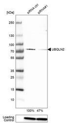
- Western blot analysis in A-431 cells transfected with control siRNA, target specific siRNA probe #1, using Anti-UBQLN2 antibody. Remaining relative intensity is presented. Loading control: Anti-GAPDH.
Supportive validation
- Submitted by
- Atlas Antibodies (provider)
- Main image

- Experimental details
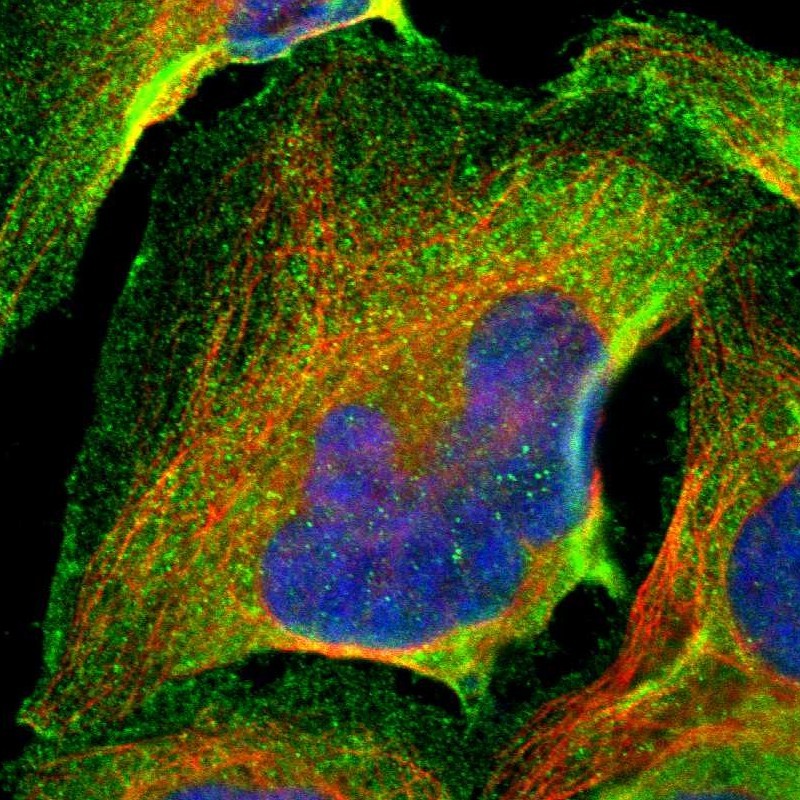
- Immunofluorescent staining of human cell line U-2 OS shows localization to plasma membrane & cytosol.
- Sample type
- HUMAN
Enhanced validation
Supportive validation
- Submitted by
- Atlas Antibodies (provider)
- Enhanced method
- Orthogonal validation
- Main image

- Experimental details
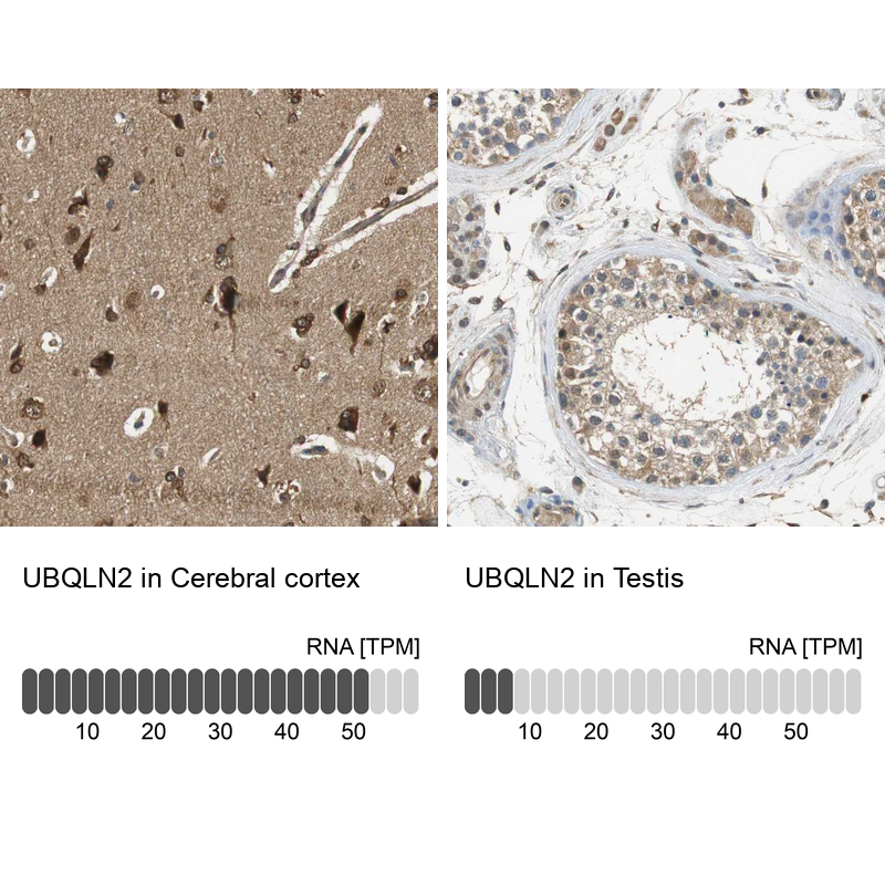
- Immunohistochemistry analysis in human cerebral cortex and testis tissues using Anti-UBQLN2 antibody. Corresponding UBQLN2 RNA-seq data are presented for the same tissues.
- Sample type
- HUMAN
Supportive validation
- Submitted by
- Atlas Antibodies (provider)
- Main image
- Experimental details
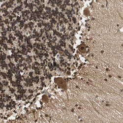
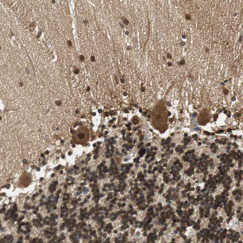
- Immunohistochemical staining of human cerebellum shows strong nuclear and cytoplasmic positivity in Purkinje cells, cells in molecular layer and cells in granular layer.
- Submitted by
- Atlas Antibodies (provider)
- Main image
- Experimental details
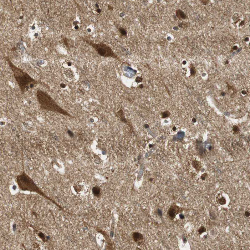
- Immunohistochemical staining of human cerebral cortex shows high expression.
- Sample type
- HUMAN

- Submitted by
- Atlas Antibodies (provider)
- Main image
- Experimental details
- Immunohistochemical staining of human testis shows low expression as expected.
- Sample type
- HUMAN

- Submitted by
- Atlas Antibodies (provider)
- Main image
- Experimental details
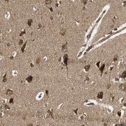
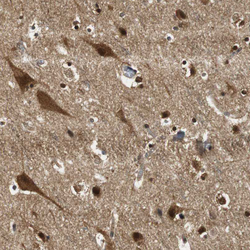
- Immunohistochemical staining of human cerebral cortex shows moderate to strong cytoplasmic positivity in neurons and glial cells.
- Sample type
- HUMAN
- Submitted by
- Atlas Antibodies (provider)
- Main image

- Experimental details
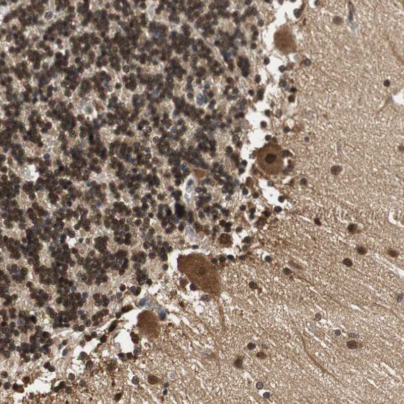
- Immunohistochemical staining of human cerebellum shows moderate to strong cytoplasmic and nuclear positivity in Purkinje cells.
- Sample type
- HUMAN
- Submitted by
- Atlas Antibodies (provider)
- Main image

- Experimental details
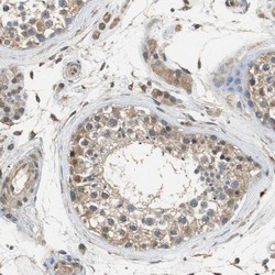
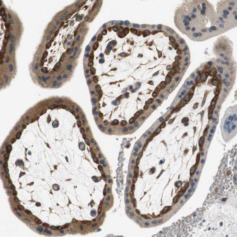
- Immunohistochemical staining of human placenta shows moderate to strong cytoplasmic positivity in cytotrophoblast.
- Sample type
- HUMAN
- Submitted by
- Atlas Antibodies (provider)
- Main image
- Experimental details
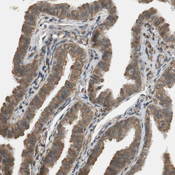
- Immunohistochemical staining of human fallopian tube shows weak to moderate cytoplasmic positivity in glandular cells.
- Sample type
- HUMAN
