Explore
 Validate
 Learn
Learn Western blot
 Chromatin Immunoprecipitation
Antibody data
- Antibody Data
- Antigen structure
- References [0]
- Comments [0]
- Validations
- Western blot [2]
- Immunocytochemistry [1]
- Immunohistochemistry [6]
- Other assay [1]
Submit
Validation data
Reference
Comment
Report error
- Product number
- UM500091 - Provider product page

- Provider
- Invitrogen Antibodies
- Product name
- IDO1 Monoclonal Antibody (UMAB126), UltraMAB™
- Antibody type
- Monoclonal
- Antigen
- Recombinant full-length protein
- Reactivity
- Human
- Host
- Mouse
- Isotype
- IgG
- Antibody clone number
- UMAB126
- Vial size
- 100 µL
- Concentration
- 0.5-1.0 mg/mL
- Storage
- -20° C, Avoid Freeze/Thaw Cycles
No comments: Submit comment
Supportive validation
- Submitted by
- Invitrogen Antibodies (provider)
- Main image
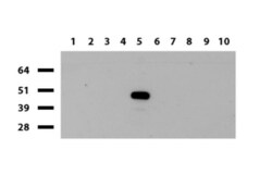
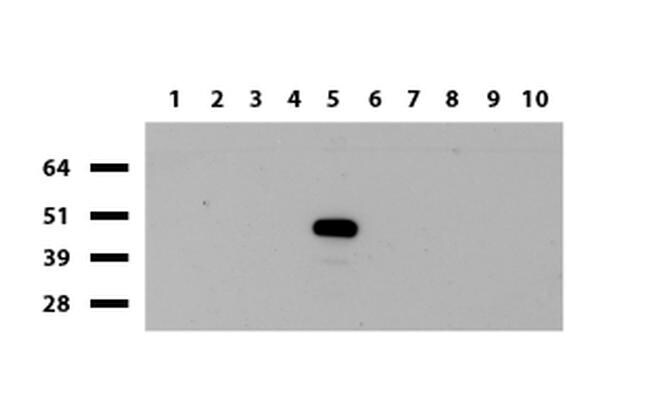
- Experimental details
- Western blot of human tissue lysates (15 µg) from 10 different tissues (1: Testis, 2: Omentum, 3: Uterus, 4: Breast, 5: Brain, 6: Thyroid, 7: Colon, 8: Spleen 9: Liver, 10: Ovary). Dilution: 1:500.
- Submitted by
- Invitrogen Antibodies (provider)
- Main image
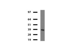
- Experimental details
- Western blot of mouse tissue lysates (20 µg) from Brian. Primary antibody dilution: 1:500. Secondary antibody dilution: Mouse TrueBlotUltra (1:1000).

Supportive validation
- Submitted by
- Invitrogen Antibodies (provider)
- Main image
- Experimental details
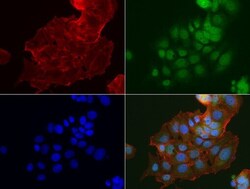
- Immunofluorescent staining of MCF-7 cells using anti-IDO1 mouse monoclonal antibody (UM500091, green, 1:100). Actin filaments were labeled with Alexa Fluor 594 Phalloidin (red), and nuclear with DAPI (blue).

Supportive validation
- Submitted by
- Invitrogen Antibodies (provider)
- Main image
- Experimental details
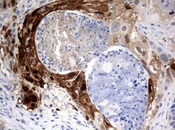
- Immunohistochemical staining of paraffin-embedded Carcinoma of Human lung tissue using anti-IDO1 mouse monoclonal antibody. (UM500091; heat-induced epitope retrieval by 10mM citric buffer, pH6.0, 120°C for 3min)

- Submitted by
- Invitrogen Antibodies (provider)
- Main image

- Experimental details
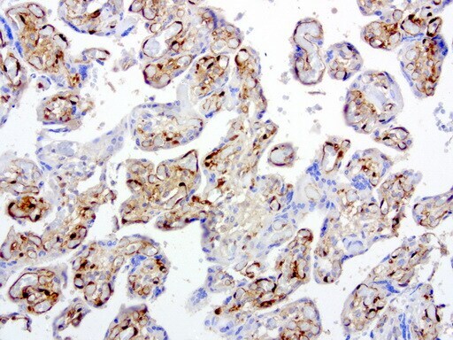
- Immunohistochemical staining of paraffin-embedded human placenta using anti-IDO1 clone UMAB126 mouse monoclonal antibody at 1:200 dilution of mg/mL using Polink2 Broad HRP DAB for detection. UM500091 requires HIER with with citrate pH6.0 at 110°C for 3 min using pressure chamber/cooker. The placenta shows strong membrane and cytoplasmic in the endothelial cells.
- Submitted by
- Invitrogen Antibodies (provider)
- Main image
- Experimental details
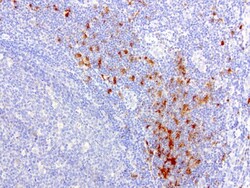
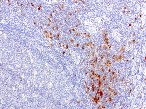
- Immunohistochemical staining of paraffin-embedded human tonsil using anti-IDO1 clone UMAB126 mouse monoclonal antibody at 1:200 dilution of mg/mL using Polink2 Broad HRP DAB for detection. UM500091 requires HIER with with citrate pH6.0 at 110°C for 3 min using pressure chamber/cooker. The tonsil shows strong membrane and cytoplasmic in the germinal center and rare strong nuclear, membrane, and cytoplasmic staining in the non germinal centers.
- Submitted by
- Invitrogen Antibodies (provider)
- Main image
- Experimental details
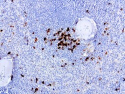
- Immunohistochemical staining of paraffin-embedded human spleen using anti-IDO1 clone UMAB126 mouse monoclonal antibody at 1:200 dilution of mg/mL using Polink2 Broad HRP DAB for detection. UM500091 requires HIER with with citrate pH6.0 at 110°C for 3 min using pressure chamber/cooker. The spleen shows very few cells staining in the red pulp with strong nuclear, membrane, and cytoplasmic staining.
- Submitted by
- Invitrogen Antibodies (provider)
- Main image

- Experimental details
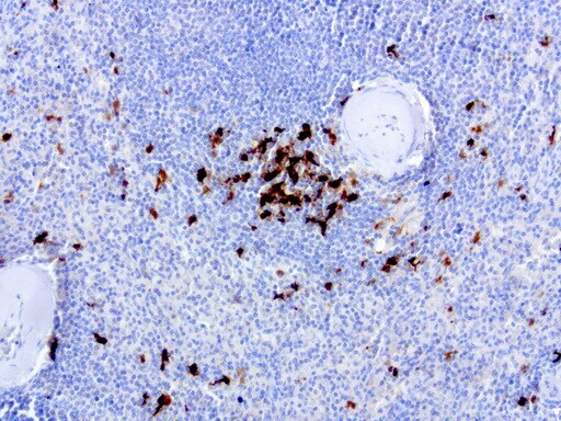
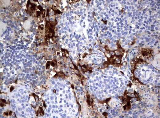
- Immunohistochemical staining of paraffin-embedded human lymph node tissue using anti-IDO1 mouse monoclonal antibody. (UM500091; heat-induced epitope retrieval by 10mM citric buffer, pH6.0, 120°C for 3min)
- Submitted by
- Invitrogen Antibodies (provider)
- Main image

- Experimental details
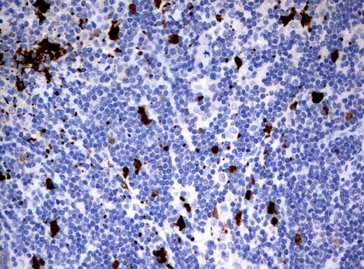
- Immunohistochemical staining of paraffin-embedded human tonsil using anti-IDO1 mouse monoclonal antibody. (UM500091; heat-induced epitope retrieval by 10mM citric buffer, pH6.0, 120°C for 3min)
Supportive validation
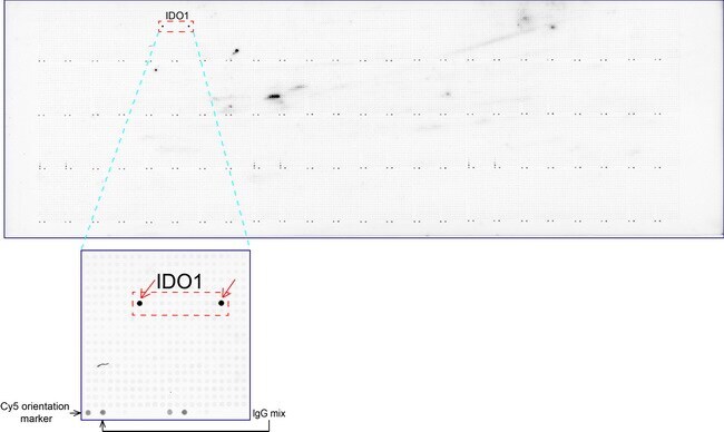
- Submitted by
- Invitrogen Antibodies (provider)
- Main image
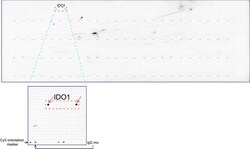
- Experimental details
- OriGene overexpression protein microarray chip was immunostained with UltraMAB anti-IDO1 mouse monoclonal antibody (UM500091). The positive reactive proteins are highlighted with two red arrows in the enlarged subarray. All the positive controls spotted in this subarray are also labeled for clarification.