 Explore
 Validate
 Learn
Learn Immunocytochemistry
Antibody data
- Antibody Data
- Antigen structure
- References [1]
- Comments [0]
- Validations
- Immunocytochemistry [1]
- Flow cytometry [2]
Submit
Validation data
Reference
Comment
Report error
- Product number
- MAB6030 - Provider product page

- Provider
- R&D Systems
- Product name
- Human Indoleamine 2,3-dioxygenase/IDO Antibody
- Antibody type
- Monoclonal
- Description
- Protein A or G purified from hybridoma culture supernatant. Detects human Indoleamine 2,3-dioxygenase/IDO in direct ELISA.
- Reactivity
- Human
- Host
- Mouse
- Conjugate
- Unconjugated
- Antigen sequence
P14902- Isotype
- IgG
- Antibody clone number
- 700838
- Vial size
- 100 ug
- Concentration
- LYOPH
- Storage
- Use a manual defrost freezer and avoid repeated freeze-thaw cycles. 12 months from date of receipt, -20 to -70 °C as supplied. 1 month, 2 to 8 °C under sterile conditions after reconstitution. 6 months, -20 to -70 °C under sterile conditions after reconstitution.
Submitted references Indoleamine 2,3-dioxygenase 1 (IDO1) activity correlates with immune system abnormalities in multiple myeloma.
Bonanno G, Mariotti A, Procoli A, Folgiero V, Natale D, De Rosa L, Majolino I, Novarese L, Rocci A, Gambella M, Ciciarello M, Scambia G, Palumbo A, Locatelli F, De Cristofaro R, Rutella S
Journal of translational medicine 2012 Dec 11;10:247
Journal of translational medicine 2012 Dec 11;10:247
No comments: Submit comment
Supportive validation
- Submitted by
- R&D Systems (provider)
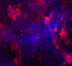
- Main image

- Experimental details
- Indoleamine 2,3-dioxygenase/IDO in A431 Human Cell Line. Indoleamine 2,3-dioxygenase/IDO was detected in immersion fixed A431 human epithelial carcinoma cells stimulated with 0.5 ng/mL of Recombinant Human IFN-gamma (Catalog # 285-IF) using Mouse Anti-Human Indoleamine 2,3-dioxygenase/IDO Monoclonal Antibody (Catalog # MAB6030) at 10 µg/mL for 3 hours at room temperature. Cells were stained using the NorthernLights™ 557-conjugated Anti-Mouse IgG Secondary Antibody (red; Catalog # NL007) and counter-stained with DAPI (blue). Specific staining was localized to cytoplasm. View our protocol for Fluorescent ICC Staining of Cells on Coverslips.
Supportive validation
- Submitted by
- R&D Systems (provider)
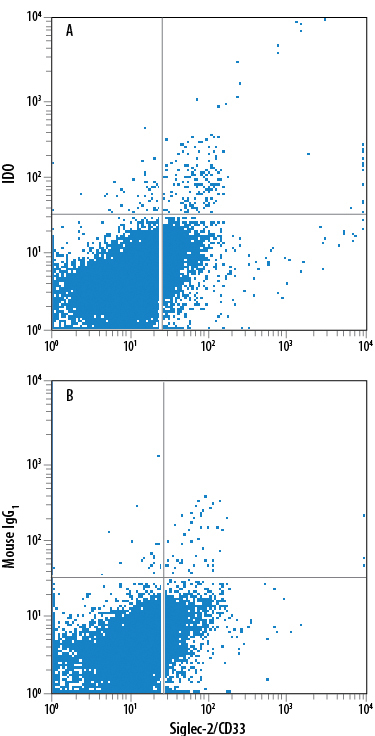
- Main image

- Experimental details
- Detection of Indoleamine 2,3-dioxygenase/IDO in Human MDSCs by Flow Cytometry. Human PBMC-derived myeloid-derived suppressor cells (MDSCs) treated with 10 ng/mL Recombinant Human IL-6 (Catalog # 206-IL) and 10 ng/mL Recombinant Human GM-CSF (Catalog # 215-GM) for 7 days were stained with Mouse Anti-Human Siglec-3/CD33 APC-conjugated Monoclonal Antibody (Catalog # FAB1137A) and either (A) Mouse Anti-Human Indoleamine 2,3-dioxygenase/IDO Monoclonal Antibody (Catalog # MAB6030) or (B) Mouse IgG1 Isotype Control (Catalog # MAB002) followed by Phycoerythrin-conjugated Anti-Mouse IgG Secondary Antibody (Catalog # F0102B). To facilitate intracellular staining, cells were fixed with paraformaldehyde and permeabilized with saponin. View our protocol for Staining Intracellular Molecules.
- Submitted by
- R&D Systems (provider)
- Main image

- Experimental details
- Detection of Indoleamine 2,3-dioxygenase/IDO in Human Monocytes by Flow Cytometry. Human Monocytes were selected from PBMC using MagCellect Human CD14+ Cell Isolation Kit (Catalog # MAGH105) and cultured overnight with (A) 50 ng/mL Recombinant Human MCSF (Catalog # 216-MC), 50 ng/mL Recombinant Human IFNg (Catalog # 285-IF) and 50 ng/mL LPS, or (B) Recombinant Human MCSF alone. Cells were stained with Mouse Anti-Human Indoleamine 2,3-dioxygenase/IDO Monoclonal Antibody (Catalog # MAB6030) followed by APC-conjugated Anti-Mouse IgG Secondary Antibody (Catalog # F0101B) and Mouse Anti-Human CD14 PE-conjugated Monoclonal Antibody (Catalog # FAB3832P). Quadrant markers were set based on Mouse IgG1 Isotype Control (Catalog # MAB002). To facilitate intracellular staining, cells were fixed with 1% paraformaldehyde and permeabilized with saponin. View our protocol for Staining Intracellular Molecules.
