Explore
 Validate
 Learn
Learn Western blot
Antibody data
- Antibody Data
- Antigen structure
- References [1]
- Comments [0]
- Validations
- Western blot [3]
- Immunohistochemistry [1]
Submit
Validation data
Reference
Comment
Report error
- Product number
- PA5-77432 - Provider product page

- Provider
- Invitrogen Antibodies
- Product name
- VGLUT3 Polyclonal Antibody
- Antibody type
- Polyclonal
- Antigen
- Synthetic peptide
- Description
- For reconstitution, we recommend adding 100 µL distilled water to a final antibody concentration of about 1 mg/mL. To use this carrier-free antibody for conjugation experiments, we strongly recommend performing another round of desalting. (Zeba Spin Desalting Columns, 7KMWCO, 0.5 mL, Product # 89882)
- Reactivity
- Human, Mouse, Rat
- Host
- Rabbit
- Isotype
- IgG
- Vial size
- 50 µL
- Concentration
- 0.8 mg/mL
- Storage
- -20°C
Submitted references Whole-brain connectivity atlas of glutamatergic and GABAergic neurons in the mouse dorsal and median raphe nuclei.
Xu Z, Feng Z, Zhao M, Sun Q, Deng L, Jia X, Jiang T, Luo P, Chen W, Tudi A, Yuan J, Li X, Gong H, Luo Q, Li A
eLife 2021 Nov 18;10
eLife 2021 Nov 18;10
No comments: Submit comment
Supportive validation
- Submitted by
- Invitrogen Antibodies (provider)
- Main image

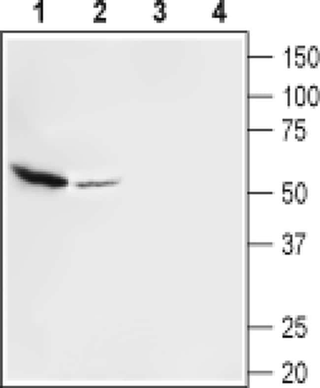
- Experimental details
- Western blot analysis of mouse brain membranes (lanes 1 and 3) and rat brain membranes (lanes 2 and 4) with VGLUT3 polyclonal antibody (Product # PA5-77432) using a dilution of 1:500.

- Submitted by
- Invitrogen Antibodies (provider)
- Main image

- Experimental details
- Western blot analysis of mouse brain membranes (lanes 1 and 3) and rat brain membranes (lanes 2 and 4) with VGLUT3 polyclonal antibody (Product # PA5-77432) using a dilution of 1:500.
- Submitted by
- Invitrogen Antibodies (provider)
- Main image
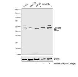
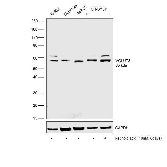
- Experimental details
- Western blot was performed using Anti-VGLUT3 Polyclonal clonal Antibody (Product # PA5-77432) and 65 kDa, 59 kDa bands corresponding to VGLUT3 were observed across cell lines tested and was increased upon Retinoic acid treatment. Whole cell Extracts (30 µg lysate) of K-562 (Lane 1), Neuro-2a (Lane 2), IMR-32 (Lane 3), SH-SY5Y (Lane 4) and SH-SY5Y treated with Retinoic Acid (10nM for 8 Days) (Lane 5) were electrophoresed using NuPAGE™ 4-12% Bis-Tris Protein Gel (Product # NP0322BOX). Resolved proteins were then transferred onto a nitrocellulose membrane (Product # IB23001) by iBlot® 2 Dry Blotting System (Product # IB21001). The blot was probed with the primary antibody (1:500 Dilution) and detected by chemiluminescence with Goat anti-Rabbit IgG (H+L), Superclonal™ Recombinant Secondary Antibody, HRP (Product # A27036, 1:4000 dilution) using the iBright FL 1000 (Product # A32752). Chemiluminescent detection was performed SuperSignal™ West Dura Extended Duration Substrate (Product # 34076)
Supportive validation
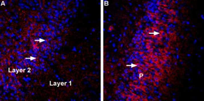
- Submitted by
- Invitrogen Antibodies (provider)
- Main image

- Experimental details
- Immunohistochemistry analysis of VGLUT3 in perfusion-fixed, frozen rat and mouse brain. Samples were probed with VGLUT3 polyclonal antibody (Product # PA5-77432) using a dilution of 1:400, and incubated with anti-rabbit-Cy3 and DAPI. A) Stained rat piriform cortex. B) Stained hippocampal CA3 region.