 Explore
 Validate
 Learn
Learn Western blot
Antibody data
- Antibody Data
- Antigen structure
- References [0]
- Comments [0]
- Validations
- Western blot [1]
- Immunohistochemistry [9]
Submit
Validation data
Reference
Comment
Report error
- Product number
- STJ13100512 - Provider product page

- Provider
- St John's Laboratory
- Product name
- Anti-VGluT3 antibody (STJ13100512)
- Antibody type
- Polyclonal
- Description
- Nz White Rabbit polyclonal antibody anti-VGluT3 is suitable for use in Immunohistochemistry and Western Blot research applications.
- Reactivity
- Human, Mouse, Rat
- Conjugate
- Unconjugated
- Antigen sequence
NA- Epitope
- NA
- Isotype
- IgG
- Antibody clone number
- NA
- Vial size
- NA
- Concentration
- NA
- Storage
- Maintain the lyophilised/reconstituted antibodies frozen at-20°C for long term storage and refrigerated at 2-8°C for a shorter term. When reconstituting, glycerol (1:1) may be added for an additional stability. Avoid freeze and thaw cycles.
- Handling
- NA
No comments: Submit comment
Supportive validation
- Submitted by
- St John's Laboratory (provider)
- Main image
- Experimental details
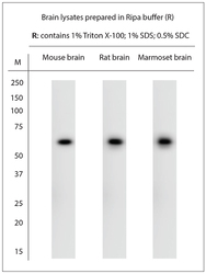
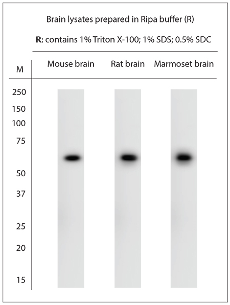
- Western blot on brain lysates prepared in Ripa buffer (critical). Blocking: 1% LFDM for 30 min at RT; primary antibody: dilution 1:500 incubated overnight at 4C.
- Sample type
- NA
- Validation comment
- NA
- Primary Ab dilution
- NA
- Other comments
- NA
- Secondary Ab
- NA
- Secondary Ab dilution
- NA
- Protocol
- NA
Supportive validation
Supportive validation
Supportive validation
Supportive validation
Supportive validation
Supportive validation
Supportive validation
Supportive validation
Supportive validation
- Submitted by
- St John's Laboratory (provider)
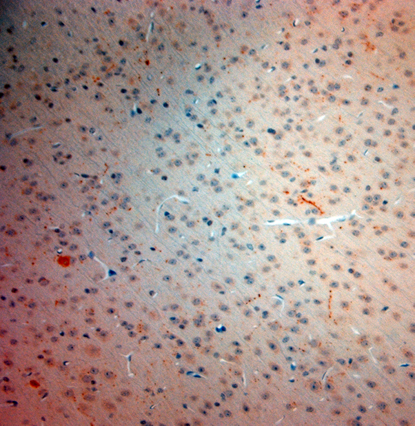
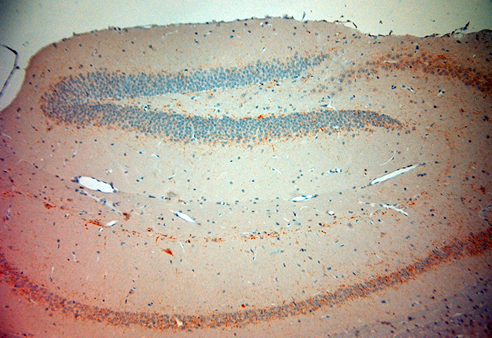
- Main image

- Experimental details
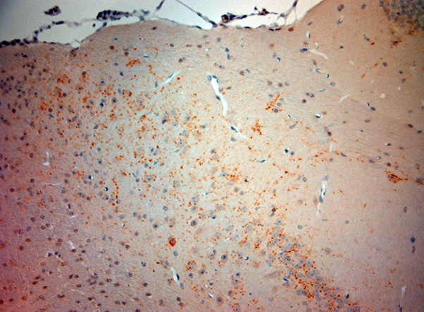
- Immunohistochemistry on paraffin sections of mouse hippocampus. The animal was perfused using Autoperfuser at a pressure of 130 mmHg with 300 ml 4% FA before being processed for paraffin embedding. HIER: Tris-EDTA, pH 9 for 20 min using Thermo PT Module. Blocking:0. 2% LFDM in TBST filtered thru 0. 2 µm. Detection was done using Novolink HRP polymer from Leica following manufacturers instructions; DAB chromogen: Candela DAB chromogen. Primary antibody: dilution 1:250, incubated 30 min at RT using Autostainer. Sections were counterstained with Harris Hematoxylin.
- Sample type
- NA
- Validation comment
- NA
- Primary Ab dilution
- NA
- Other comments
- NA
- Secondary Ab
- NA
- Secondary Ab dilution
- NA
- Protocol
- NA
Supportive validation
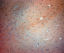
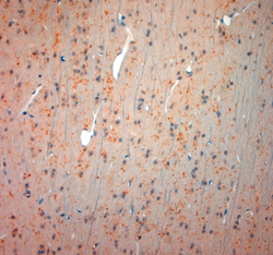
- Submitted by
- St John's Laboratory (provider)
- Main image

- Experimental details
- Immunohistochemistry on paraffin sections of mouse hippocampus. The animal was perfused using Autoperfuser at a pressure of 130 mmHg with 300 ml 4% FA before being processed for paraffin embedding. HIER: Tris-EDTA, pH 9 for 20 min using Thermo PT Module. Blocking:0. 2% LFDM in TBST filtered thru 0. 2 µm. Detection was done using Novolink HRP polymer from Leica following manufacturers instructions; DAB chromogen: Candela DAB chromogen. Primary antibody: dilution 1:250, incubated 30 min at RT using Autostainer. Sections were counterstained with Harris Hematoxylin.
- Sample type
- NA
- Validation comment
- NA
- Primary Ab dilution
- NA
- Other comments
- NA
- Secondary Ab
- NA
- Secondary Ab dilution
- NA
- Protocol
- NA
Supportive validation
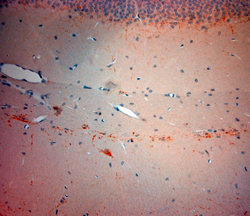
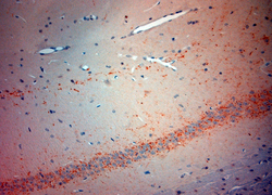
- Submitted by
- St John's Laboratory (provider)
- Main image
- Experimental details
- Immunohistochemistry on paraffin sections of mouse hippocampus. The animal was perfused using Autoperfuser at a pressure of 130 mmHg with 300 ml 4% FA before being processed for paraffin embedding. HIER: Tris-EDTA, pH 9 for 20 min using Thermo PT Module. Blocking:0. 2% LFDM in TBST filtered thru 0. 2 µm. Detection was done using Novolink HRP polymer from Leica following manufacturers instructions; DAB chromogen: Candela DAB chromogen. Primary antibody: dilution 1:250, incubated 30 min at RT using Autostainer. Sections were counterstained with Harris Hematoxylin.
- Sample type
- NA
- Validation comment
- NA
- Primary Ab dilution
- NA
- Other comments
- NA
- Secondary Ab
- NA
- Secondary Ab dilution
- NA
- Protocol
- NA

Supportive validation
- Submitted by
- St John's Laboratory (provider)
- Main image

- Experimental details
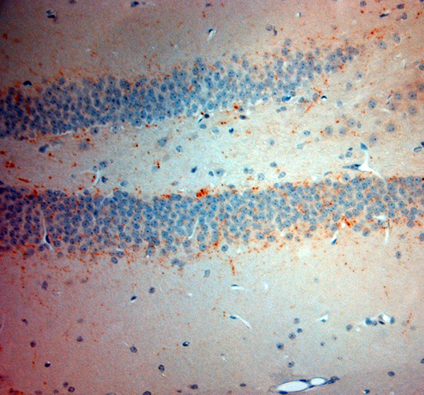
- Immunohistochemistry on paraffin sections of mouse brain (hippocampus). The animal was perfused using Autoperfuser at a pressure of 130 mmHg with 300 ml 4% FA before being processed for paraffin embedding. HIER: Tris-EDTA, pH 9 for 20 min using Thermo PT Module. Blocking:0. 2% LFDM in TBST filtered thru 0. 2 µm. Detection was done using Novolink HRP polymer from Leica following manufacturers instructions; DAB chromogen: Candela DAB chromogen. Primary antibody: dilution 1:250, incubated 30 min at RT using Autostainer. Sections were counterstained with Harris Hematoxylin.
- Sample type
- NA
- Validation comment
- NA
- Primary Ab dilution
- NA
- Other comments
- NA
- Secondary Ab
- NA
- Secondary Ab dilution
- NA
- Protocol
- NA
Supportive validation
- Submitted by
- St John's Laboratory (provider)
- Main image
- Experimental details
- Immunohistochemistry on paraffin sections of mouse brain. The animal was perfused using Autoperfuser at a pressure of 130 mmHg with 300 ml 4% FA before being processed for paraffin embedding. HIER: Tris-EDTA, pH 9 for 20 min using Thermo PT Module. Blocking:0. 2% LFDM in TBST filtered thru 0. 2 µm. Detection was done using Novolink HRP polymer from Leica following manufacturers instructions; DAB chromogen: Candela DAB chromogen. Primary antibody: dilution 1:250, incubated 30 min at RT using Autostainer. Sections were counterstained with Harris Hematoxylin.
- Sample type
- NA
- Validation comment
- NA
- Primary Ab dilution
- NA
- Other comments
- NA
- Secondary Ab
- NA
- Secondary Ab dilution
- NA
- Protocol
- NA
