Explore
 Validate
 Learn
Learn Western blot
Antibody data
- Antibody Data
- Antigen structure
- References [0]
- Comments [0]
- Validations
- Western blot [3]
- Immunohistochemistry [1]
- Flow cytometry [1]
Submit
Validation data
Reference
Comment
Report error
- Product number
- TA328616 - Provider product page

- Provider
- OriGene
- Product name
- Rabbit Polyclonal Anti-Bombesin Receptor 3 (extracellular)
- Antibody type
- Polyclonal
- Description
- Rabbit Polyclonal Anti-Bombesin Receptor 3 (extracellular)
- Host
- Rabbit
- Conjugate
- Unconjugated
- Epitope
- BRS3
- Antibody clone number
- NULL
- Vial size
- 200 µl
- Concentration
- NULL
No comments: Submit comment
Supportive validation
- Submitted by
- OriGene (provider)
- Main image
- Experimental details
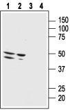
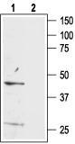
- Western blot analysis of rat brain membranes: 1. Anti-Bombesin Receptor 3 (extracellular) antibody, (1:200). 2. Anti-Bombesin Receptor 3 (extracellular) antibody, preincubated with the control peptide antigen.
- Validation comment
- WB
- Submitted by
- OriGene (provider)
- Main image

- Experimental details
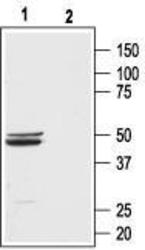
- Western blot analysis of human malignant melanoma cell line Malme-3M (lanes 1 and 3) and human normal skin fibroblast cell line Malme-3 (lanes 2 and 4): 1, 2. Anti-Bombesin Receptor 3 (extracellular) antibody, (1:500). 3, 4. Anti-Bombesin Receptor 3 (extracellular) antibody, preincubated with the control peptide antigen.
- Validation comment
- WB
- Submitted by
- OriGene (provider)
- Main image

- Experimental details
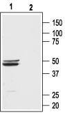
- Western blot analysis of mouse brain lysate: 1. Anti-Bombesin Receptor 3 (extracellular) antibody, (1:200). 2. Anti-Bombesin Receptor 3 (extracellular) antibody, preincubated with the control peptide antigen.
- Validation comment
- WB
Supportive validation
- Submitted by
- OriGene (provider)
- Main image

- Experimental details
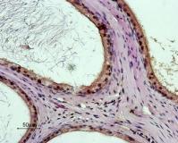
- Expression of Bombesin Receptor 3 in rat testes. Immunohistochemical staining of paraffin embedded rat testes section using Anti-Bombesin Receptor 3 (extracellular) antibody, (1:50). The area of the efferent ductules near the epididymis is shown. Intense stain (brown) is specific for the pseudostratified epithelium of the efferent ductules. DAB is used for the color reaction. H&E is used as the counterstain.
- Validation comment
- IHC
Supportive validation
- Submitted by
- OriGene (provider)
- Main image
- Experimental details
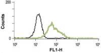
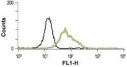
- Indirect flow cytometry analysis in human promyelocytic leukemia HL-60 cells: black line, Unstained cells + goat-anti-rabbit-FITC. green line, Cells + Anti-Bombesin Receptor 3 (extracellular) antibody, (5 ug) + goat-anti-rabbit-FITC.
- Validation comment
- FC