Explore
 Validate
 Learn
Learn Immunohistochemistry
Antibody data
- Antibody Data
- Antigen structure
- References [0]
- Comments [0]
- Validations
- Immunohistochemistry [6]
Submit
Validation data
Reference
Comment
Report error
- Product number
- AMAb91639 - Provider product page

- Provider
- Atlas Antibodies
- Product name
- Anti-PLP1
- Antibody type
- Monoclonal
- Description
- Monoclonal Antibody against Human PLP1, Clone ID: CL10622, Gene description: Proteolipid protein 1, Alternative Gene Names: GPM6C, PLP, SPG2, Validated applications: IHC, Uniprot ID: P60201, Storage: Store at +4°C for short term storage. Long time storage is recommended at -20°C.
- Reactivity
- Human, Rat
- Host
- Mouse
- Conjugate
- Unconjugated
- Isotype
- IgG
- Antibody clone number
- CL10622
- Vial size
- 100 µl
- Concentration
- 1.0 mg/ml
- Storage
- Store at +4°C for short term storage. Long time storage is recommended at -20°C.
No comments: Submit comment
Enhanced validation
Supportive validation
- Submitted by
- Atlas Antibodies (provider)
- Enhanced method
- Orthogonal validation
- Main image

- Experimental details
- Immunohistochemistry analysis in human cerebral cortex and tonsil tissues using AMAb91639 antibody. Corresponding PLP1 RNA-seq data are presented for the same tissues.
- Sample type
- Human
- Protocol
- Protocol

Supportive validation
- Submitted by
- Atlas Antibodies (provider)
- Main image

- Experimental details
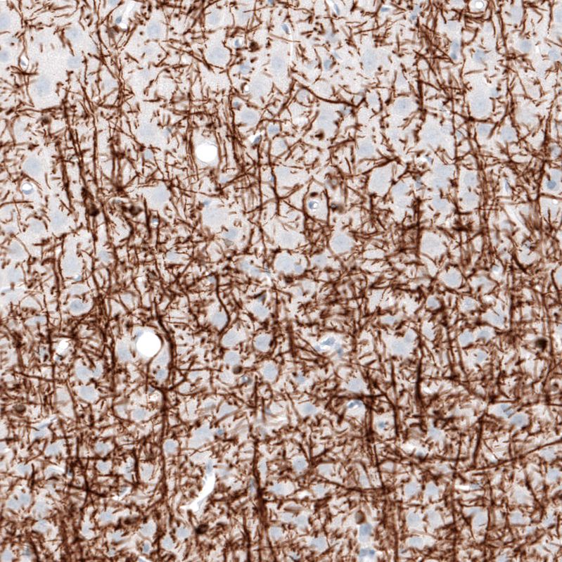
- Immunohistochemical staining of human cerebral cortex shows strong positivity in myelinated neural fibers.
- Sample type
- Human
- Protocol
- Protocol

- Submitted by
- Atlas Antibodies (provider)
- Main image

- Experimental details
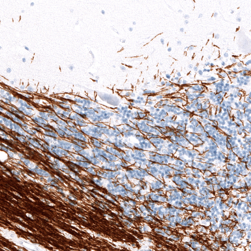
- Immunohistochemical staining of human cerebellum shows strong positivity in myelinated neuronal processes.
- Sample type
- Human
- Protocol
- Protocol
- Submitted by
- Atlas Antibodies (provider)
- Main image

- Experimental details
- Immunohistochemical staining of human kidney shows no positivity as expected.
- Sample type
- Human
- Protocol
- Protocol

- Submitted by
- Atlas Antibodies (provider)
- Main image

- Experimental details
- Immunohistochemical staining of human tonsil shows no positivity in lymphoid cells as expected.
- Sample type
- Human
- Protocol
- Protocol

- Submitted by
- Atlas Antibodies (provider)
- Main image

- Experimental details
- Immunohistochemical staining of rat cerebral cortex shows strong positivity in myelinated neuronal processes and in oligodendrocytes cell bodies.
- Sample type
- Rat
- Protocol
- Protocol