Explore
 Validate
 Learn
Learn Western blot
Antibody data
- Antibody Data
- Antigen structure
- References [0]
- Comments [0]
- Validations
- Western blot [1]
- Immunocytochemistry [1]
- Immunohistochemistry [2]
- Flow cytometry [1]
Submit
Validation data
Reference
Comment
Report error
- Product number
- BSM-52099R - Provider product page

- Provider
- Invitrogen Antibodies
- Product name
- Histone H2B Monoclonal Antibody (3A6)
- Antibody type
- Monoclonal
- Antigen
- Recombinant full-length protein
- Reactivity
- Human, Mouse, Rat
- Host
- Rabbit
- Isotype
- IgG
- Antibody clone number
- 3A6
- Vial size
- 100 µL
- Concentration
- 1 mg/mL
- Storage
- -20°C
No comments: Submit comment
Supportive validation
- Submitted by
- Invitrogen Antibodies (provider)
- Main image
- Experimental details
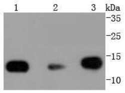
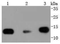
- Lane 1: Hela; Lane 2: NIH/3T3; Lane 3: PC12 lysates probed with Histone H2B (3A6) Monoclonal Antibody (bsm-52099R) at 1:1000 overnight at 4°C. Followed by a conjugated secondary antibody.
Supportive validation
- Submitted by
- Invitrogen Antibodies (provider)
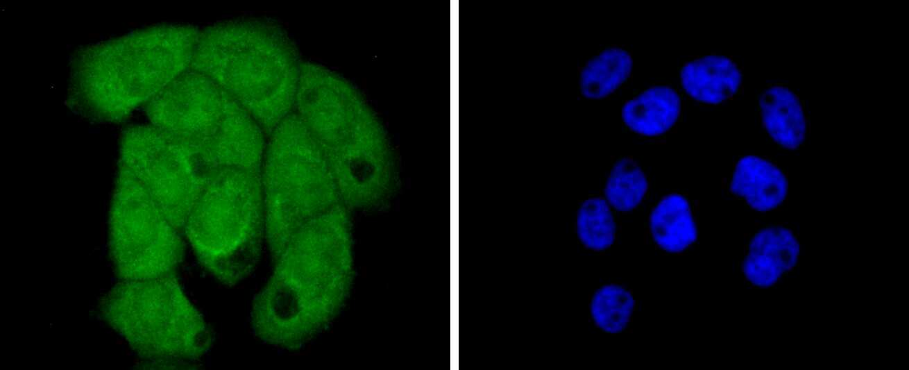
- Main image
- Experimental details
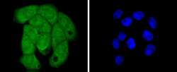
- A431 cells were stained with Histone H2B (3A6) Monoclonal Antibody (bsm-52099R) at [1:200] incubated overnight at 4C, followed by secondary antibody incubation, DAPI staining of the nuclei and detection.
Supportive validation
- Submitted by
- Invitrogen Antibodies (provider)
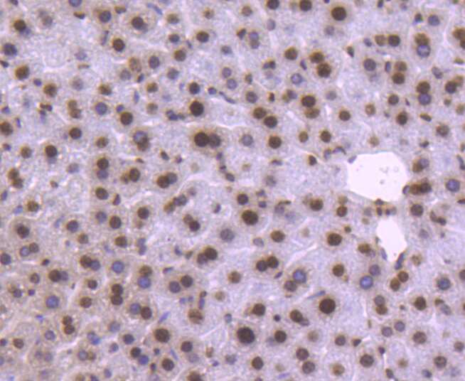
- Main image
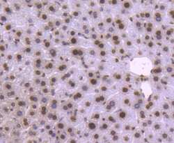
- Experimental details
- Paraformaldehyde-fixed, paraffin embedded mouse liver; Antigen retrieval by boiling in sodium citrate buffer (pH6) for 15min; Block endogenous peroxidase by 3% hydrogen peroxide for 30 minutes; Blocking buffer (normal serum) at 37°C for 20min; Antibody incubation with Histone H2B (3A6) Monoclonal Antibody (bsm-52099R) at 1:50 overnight at 4°C, followed by a conjugated secondary and DAB staining.
- Submitted by
- Invitrogen Antibodies (provider)
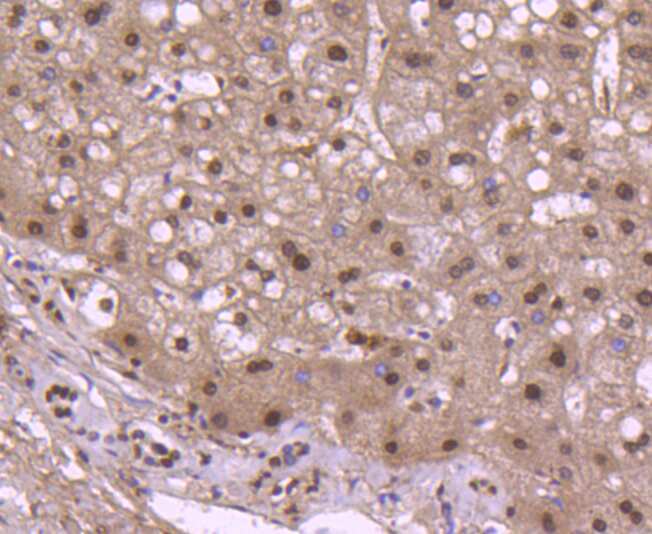
- Main image

- Experimental details
- Paraformaldehyde-fixed and paraffin-embedded Human Liver tissue incubated with Histone H2B (3A6) Monoclonal Antibody (bsm-52099R) at 1:100, overnight at 4°C, followed by a conjugated secondary antibody and DAB staining. Counterstained with hematoxylin.
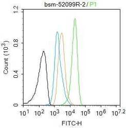
Supportive validation
- Submitted by
- Invitrogen Antibodies (provider)
- Main image
- Experimental details
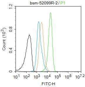
- A431 cells (black) were fixed with 4% PFA for 10 min at room temperature, permeabilized with 90% ice-cold methanol for 20 min at -20°C, and incubated in 5% BSA blocking buffer for 30 min at room temperature. Cells were then stained with Histone H2B Antibody (bsm-52099R) at 1:50 dilution in blocking buffer and incubated for 30 min at room temperature, washed twice with 2% BSA in PBS, followed by secondary antibody (blue) incubation for 40 min at room temperature. Acquisitions of 20,000 events were performed. Cells stained with primary antibody (green), and isotype control (orange).