Explore
 Validate
 Learn
Learnsc-13132
antibody from Santa Cruz Biotechnology
Targeting: HSPB1
CMT2F, Hs.76067, Hsp25, HSP27, HSP28
 Western blot
Antibody data
- Antibody Data
- Antigen structure
- References [3]
- Comments [0]
- Validations
- Western blot [1]
- Immunohistochemistry [1]
Submit
Validation data
Reference
Comment
Report error
- Product number
- sc-13132 - Provider product page

- Provider
- Santa Cruz Biotechnology
- Proper citation
- Santa Cruz Biotechnology Cat#sc-13132, RRID:AB_627755
- Product name
- Anti-HSPB1
- Antibody type
- Monoclonal
- Antigen
- Recombinant full-length protein
- Reactivity
- Human
- Host
- Mouse
Submitted references Differential expression of ANXA6, HSP27, PRDX2, NCF2, and TPM4 during uterine cervix carcinogenesis: diagnostic and prognostic value
Loss of specific chaperones involved in membrane glycoprotein biosynthesis during the maturation of human erythroid progenitor cells.
Phosphorylation of Ser78 of Hsp27 correlated with HER-2/neu status and lymph node positivity in breast cancer.
M I Lomnytska, S Becker, I Bodin, A Olsson, K Hellman, A-C Hellström, M Mints, U Hellman, G Auer, S Andersson
British Journal of Cancer 2010 Nov;104(1):110-119
British Journal of Cancer 2010 Nov;104(1):110-119
Loss of specific chaperones involved in membrane glycoprotein biosynthesis during the maturation of human erythroid progenitor cells.
Patterson ST, Li J, Kang JA, Wickrema A, Williams DB, Reithmeier RA
The Journal of biological chemistry 2009 May 22;284(21):14547-57
The Journal of biological chemistry 2009 May 22;284(21):14547-57
Phosphorylation of Ser78 of Hsp27 correlated with HER-2/neu status and lymph node positivity in breast cancer.
Zhang D, Wong LL, Koay ES
Molecular cancer 2007 Aug 14;6:52
Molecular cancer 2007 Aug 14;6:52
No comments: Submit comment
Supportive validation
- Submitted by
- per
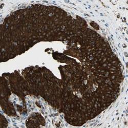
- Main image

- Experimental details
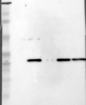
- Western blot analysis of antibody specificity using a routine panel composed of IgG/HSA-depleted human plasma and protein lysates from selected human tissues and cell lines.
- Validation comment
- Band of predicted size in kDa (+/-20%) with additional bands present.
- Primary Ab dilution
- 1:500
- Secondary Ab dilution
- 1:7000
- Lane 1
- Marker [kDa]: 220, 112, 84, 47, 32, 26, 16.8
- Lane 2
- RT-4
- Lane 3
- U-251MG sp
- Lane 4
- Human Plasma
- Lane 5
- Liver
- Lane 6
- Tonsil
- Theoretical target weight
- [kDa] 23
Supportive validation
- Submitted by
- per
- Main image
- Experimental details
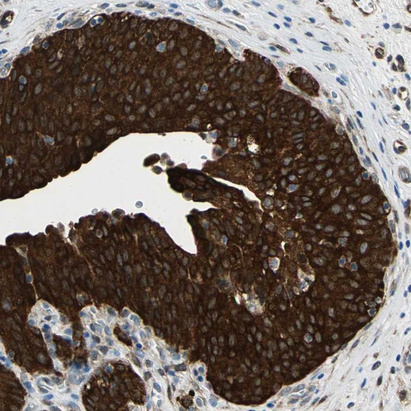
- Immunohistochemical staining of human urinary bladder shows strong cytoplasmic positivity in urothelial cells.
- Validation comment
- Two independent antibodies targeting one protein yielding similar staining patterns. Staining pattern consistent with experimental and/or bioinformatic data.