Explore
 Validate
 Learn
Learn Western blot
 Immunocytochemistry
Antibody data
- Antibody Data
- Antigen structure
- References [0]
- Comments [0]
- Validations
- Western blot [5]
- Immunocytochemistry [1]
- Immunoprecipitation [1]
- Immunohistochemistry [1]
Submit
Validation data
Reference
Comment
Report error
- Product number
- GTX112964 - Provider product page

- Provider
- GeneTex
- Proper citation
- GeneTex Cat#GTX112964, RRID:AB_11172258
- Product name
- HSP27 antibody
- Antibody type
- Polyclonal
- Reactivity
- Human
- Host
- Rabbit
No comments: Submit comment
Enhanced validation
Supportive validation
- Submitted by
- GeneTex (provider)
- Enhanced method
- Genetic validation
- Main image
- Experimental details
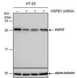
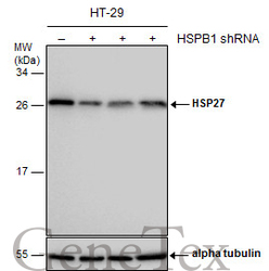
- Non-transfected (¡V) and transfected (+) HT-29 whole cell extracts (30 ?g) were separated by 12% SDS-PAGE, and the membrane was blotted with HSP27 antibody (GTX112964) diluted at 1:5000.
Supportive validation
- Submitted by
- GeneTex (provider)
- Main image
- Experimental details
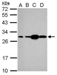
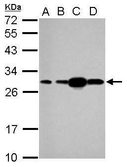
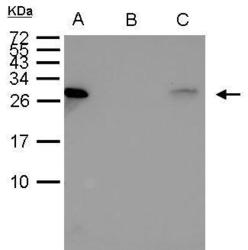
- Sample (30 ug of whole cell lysate) A: 293T B: A431 C: HeLa D: HepG2 12% SDS PAGE GTX112964 diluted at 1:1000
- Submitted by
- GeneTex (provider)
- Main image
- Experimental details
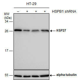
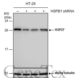
- Non-transfected (¡V) and transfected (+) HT-29 whole cell extracts (30 ?g) were separated by 12% SDS-PAGE, and the membrane was blotted with HSP27 antibody (GTX112964) diluted at 1:5000.
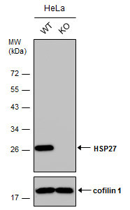
- Submitted by
- GeneTex (provider)
- Main image
- Experimental details
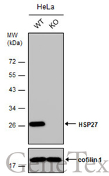
- Wild-type (WT) and HSP27 knockout (KO) HeLa cell extracts (30 ?g) were separated by 12% SDS-PAGE, and the membrane was blotted with HSP27 antibody (GTX112964) diluted at 1:20000. The HRP-conjugated anti-rabbit IgG antibody (GTX213110-01) was used to detect the primary antibody.
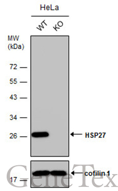
- Submitted by
- GeneTex (provider)
- Main image
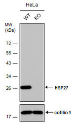
- Experimental details
- Wild-type (WT) and HSP27 knockout (KO) HeLa cell extracts (30 ?g) were separated by 12% SDS-PAGE, and the membrane was blotted with HSP27 antibody (GTX112964) diluted at 1:20000. The HRP-conjugated anti-rabbit IgG antibody (GTX213110-01) was used to detect the primary antibody.
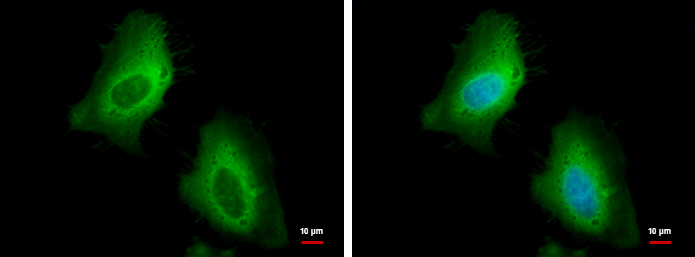
Supportive validation
- Submitted by
- GeneTex (provider)
- Main image

- Experimental details
- HSP27 antibody detects HSP27 protein at cytoplasm by immunofluorescent analysis.Sample: HeLa cells were fixed in 4% paraformaldehyde at RT for 15 min.Green: HSP27 protein stained by HSP27 antibody (GTX112964) diluted at 1:500.Blue: Hoechst 33342 staining.
Supportive validation
- Submitted by
- GeneTex (provider)
- Main image
- Experimental details
- HSP27 antibody immunoprecipitates HSP27 protein in IP experiments. IP Sample: 1000 ?g HeLa whole cell lysate/extract A. 40 £gg HeLa whole cell lysate/extract B. Control with 2.5 £gg of preimmune rabbit IgG C. Immunoprecipitation of HSP27 protein by 2.5 £gg of HSP27 (GTX112964) 15% SDS-PAGE The immunoprecipitated HSP27 protein was detected by HSP27 antibody (GTX112964) diluted at 1:1000. EasyBlot anti-rabbit IgG (GTX221666-01) was used as a secondary reagent.

Supportive validation
- Submitted by
- GeneTex (provider)
- Main image
- Experimental details
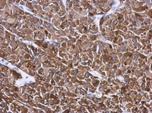
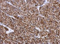
- Immunohistochemical analysis of paraffin-embedded U87 xenograft, using HSP27(GTX112964) antibody at 1:500 dilution.