MA3-015
antibody from Invitrogen Antibodies
Targeting: HSPB1
CMT2F, Hs.76067, Hsp25, HSP27, HSP28
 Western blot
 ELISA
ELISA Immunocytochemistry
 Immunoprecipitation
 Immunohistochemistry
 Immunoelectron microscopy
 Other assay
Antibody data
- Antibody Data
- Antigen structure
- References [39]
- Comments [0]
- Validations
- Western blot [3]
- Immunocytochemistry [5]
- Immunohistochemistry [3]
- Other assay [2]
Submit
Validation data
Reference
Comment
Report error
- Product number
- MA3-015 - Provider product page

- Provider
- Invitrogen Antibodies
- Product name
- HSP27 Monoclonal Antibody (G3.1)
- Antibody type
- Monoclonal
- Antigen
- Purifed from natural sources
- Description
- MA3-015 detects heat shock protein 27 kDa (HSP27) from human, rat, mouse, monkey, and canine samples. MA3-015 has been successfully used in Western blot, immunohistochemistry (paraffin), immunoprecipitation, immunofluorescence, immunocytochemistry and ELISA procedures. By Western blot, this antibody detects an ~24 kDa protein representing HSP27 in MCF-7 cell extract. Immunohistochemical staining of human cervix with MA3-015 results in intense cytoplasmic staining of the epithelium within the parabasal cell layer. Western blot detection of HSP27 in canine heart samples is weak and may require optimization. The MA3-015 antigen is partially purified HSP27 derived from MCF-7 cytosol. The mouse HSP27 sequence is strongly conserved within the human sequence, so cross-reactivity with mouse HSP27 is expected. Antibodies to this protein (and modification) were previously sold as part of a Thermo Scientific Cellomics High Content Screening Kit. This replacement antibody is now recommended for researchers who need an antibody for high content cell based assays. It has been thoroughly tested and validated for cellular immunofluorescence (IF) applications. Further optimization including the selection of the most appropriate fluorescent Dylight conjugated secondary antibody may have to be performed for your high content assay.
- Reactivity
- Human, Rat, Canine
- Host
- Mouse
- Isotype
- IgG
- Antibody clone number
- G3.1
- Vial size
- 100 µL
- Concentration
- Conc. Not Determined
- Storage
- -20° C, Avoid Freeze/Thaw Cycles
Submitted references Cyanidin 3-O-arabinoside suppresses DHT-induced dermal papilla cell senescence by modulating p38-dependent ER-mitochondria contacts.
Constitutively active androgen receptor supports the metastatic phenotype of endocrine-resistant hormone receptor-positive breast cancer.
Comp34 displays potent preclinical antitumor efficacy in triple-negative breast cancer via inhibition of NUDT3-AS4, a novel oncogenic long noncoding RNA.
Increased FXYD1 and PGC-1α mRNA after blood flow-restricted running is related to fibre type-specific AMPK signalling and oxidative stress in human muscle.
Small heat shock proteins translocate to the cytoskeleton in human skeletal muscle following eccentric exercise independently of phosphorylation.
Altered expression of HSP27 and HSP70 in distal oesophageal mucosa in patients with gastro-oesophageal reflux disease subjected to fundoplication.
Small-molecule proteostasis regulators for protein conformational diseases.
Spatiotemporal expression of Hsp20 and its phosphorylation in hippocampal CA1 pyramidal neurons after transient forebrain ischemia.
[The effect of resistance-related proteins on the prognosis and survival of patients with osteosarcoma: an immunohistochemical analysis].
Frequent PTEN genomic alterations and activated phosphatidylinositol 3-kinase pathway in basal-like breast cancer cells.
fMLP induces Hsp27 expression, attenuates NF-kappaB activation, and confers intestinal epithelial cell protection.
Identification of 14-3-3sigma as a contributor to drug resistance in human breast cancer cells using functional proteomic analysis.
Expressions of HSP70 and HSP27 in hepatocellular carcinoma.
Expressions of HSP70 and HSP27 in hepatocellular carcinoma.
The c-Yes 3'-UTR contains adenine/uridine-rich elements that bind AUF1 and HuR involved in mRNA decay in breast cancer cells.
Electromagnetic fields at mobile phone frequency induce apoptosis and inactivation of the multi-chaperone complex in human epidermoid cancer cells.
Immunohistochemical study of heat shock proteins 27, 60 and 70 in the normal human adrenal and in adrenal tumors with suppressed ACTH production.
Down-regulation of heat shock protein 27 in neuronal cells and non-neuronal cells expressing mutant ataxin-3.
Expression of heat-shock proteins hsp27, hsp70 and hsp90 in malignant epithelial tumour of the ovaries.
Expression of heat-shock proteins hsp27, hsp70 and hsp90 in malignant epithelial tumour of the ovaries.
Heat shock protein 27 prevents cellular polyglutamine toxicity and suppresses the increase of reactive oxygen species caused by huntingtin.
Differential proteome analysis of replicative senescence in rat embryo fibroblasts.
Differential proteome analysis of replicative senescence in rat embryo fibroblasts.
Regulation of human CLC-3 channels by multifunctional Ca2+/calmodulin-dependent protein kinase.
Polyglutamine expansions cause decreased CRE-mediated transcription and early gene expression changes prior to cell death in an inducible cell model of Huntington's disease.
Polyglutamine expansions cause decreased CRE-mediated transcription and early gene expression changes prior to cell death in an inducible cell model of Huntington's disease.
The Fas-induced apoptosis analyzed by high throughput proteome analysis.
Differential expression of Hsp27 in normal oesophagus, Barrett's metaplasia and oesophageal adenocarcinomas.
Heat shock proteins hsp27 and hsp70: lack of correlation with response to tamoxifen and clinical course of disease in estrogen receptor-positive metastatic breast cancer (a Southwest Oncology Group Study).
Expression of heat shock proteins in squamous cell carcinoma of the tongue: an immunohistochemical study.
Expression of heat shock proteins in squamous cell carcinoma of the tongue: an immunohistochemical study.
Chromosomal assignments of human 27-kDa heat shock protein gene family.
Chromosomal assignments of human 27-kDa heat shock protein gene family.
A cDNA for the estradiol-regulated 24K protein: control of mRNA levels in MCF-7 cells.
A cDNA for the estradiol-regulated 24K protein: control of mRNA levels in MCF-7 cells.
A new marker of maturation in the cervix: the estrogen-regulated 24K protein.
Quantitative enzyme-linked immunosorbent assay for the estrogen-regulated Mr 24,000 protein in human breast tumors: correlation with estrogen and progesterone receptors.
Evidence for modulation of a 24K protein in human endometrium during the menstrual cycle.
Detection of a Mr 24,000 estrogen-regulated protein in human breast cancer by monoclonal antibodies.
Jung YH, Chae CW, Choi GE, Shin HC, Lim JR, Chang HS, Park J, Cho JH, Park MR, Lee HJ, Han HJ
Journal of biomedical science 2022 Mar 7;29(1):17
Journal of biomedical science 2022 Mar 7;29(1):17
Constitutively active androgen receptor supports the metastatic phenotype of endocrine-resistant hormone receptor-positive breast cancer.
Bahnassy S, Thangavel H, Quttina M, Khan AF, Dhanyalayam D, Ritho J, Karami S, Ren J, Bawa-Khalfe T
Cell communication and signaling : CCS 2020 Sep 18;18(1):154
Cell communication and signaling : CCS 2020 Sep 18;18(1):154
Comp34 displays potent preclinical antitumor efficacy in triple-negative breast cancer via inhibition of NUDT3-AS4, a novel oncogenic long noncoding RNA.
Hao Q, Wang P, Dutta P, Chung S, Li Q, Wang K, Li J, Cao W, Deng W, Geng Q, Schrode K, Shaheen M, Wu K, Zhu D, Chen QH, Chen G, Elshimali Y, Vadgama J, Wu Y
Cell death & disease 2020 Dec 11;11(12):1052
Cell death & disease 2020 Dec 11;11(12):1052
Increased FXYD1 and PGC-1α mRNA after blood flow-restricted running is related to fibre type-specific AMPK signalling and oxidative stress in human muscle.
Christiansen D, Murphy RM, Bangsbo J, Stathis CG, Bishop DJ
Acta physiologica (Oxford, England) 2018 Jun;223(2):e13045
Acta physiologica (Oxford, England) 2018 Jun;223(2):e13045
Small heat shock proteins translocate to the cytoskeleton in human skeletal muscle following eccentric exercise independently of phosphorylation.
Frankenberg NT, Lamb GD, Overgaard K, Murphy RM, Vissing K
Journal of applied physiology (Bethesda, Md. : 1985) 2014 Jun 1;116(11):1463-72
Journal of applied physiology (Bethesda, Md. : 1985) 2014 Jun 1;116(11):1463-72
Altered expression of HSP27 and HSP70 in distal oesophageal mucosa in patients with gastro-oesophageal reflux disease subjected to fundoplication.
Rantanen T, Honkanen T, Paavonen T, Rantanen L, Oksala N
European journal of surgical oncology : the journal of the European Society of Surgical Oncology and the British Association of Surgical Oncology 2011 Feb;37(2):168-74
European journal of surgical oncology : the journal of the European Society of Surgical Oncology and the British Association of Surgical Oncology 2011 Feb;37(2):168-74
Small-molecule proteostasis regulators for protein conformational diseases.
Calamini B, Silva MC, Madoux F, Hutt DM, Khanna S, Chalfant MA, Saldanha SA, Hodder P, Tait BD, Garza D, Balch WE, Morimoto RI
Nature chemical biology 2011 Dec 25;8(2):185-96
Nature chemical biology 2011 Dec 25;8(2):185-96
Spatiotemporal expression of Hsp20 and its phosphorylation in hippocampal CA1 pyramidal neurons after transient forebrain ischemia.
Niwa M, Hara A, Taguchi A, Aoki H, Kozawa O, Mori H
Neurological research 2009 Sep;31(7):721-7
Neurological research 2009 Sep;31(7):721-7
[The effect of resistance-related proteins on the prognosis and survival of patients with osteosarcoma: an immunohistochemical analysis].
Ozger H, Eralp L, Atalar AC, Toker B, Esberk Ateş L, Sungur M, Bilgiç B, Ayan I
Acta orthopaedica et traumatologica turcica 2009 Jan-Feb;43(1):28-34
Acta orthopaedica et traumatologica turcica 2009 Jan-Feb;43(1):28-34
Frequent PTEN genomic alterations and activated phosphatidylinositol 3-kinase pathway in basal-like breast cancer cells.
Marty B, Maire V, Gravier E, Rigaill G, Vincent-Salomon A, Kappler M, Lebigot I, Djelti F, Tourdès A, Gestraud P, Hupé P, Barillot E, Cruzalegui F, Tucker GC, Stern MH, Thiery JP, Hickman JA, Dubois T
Breast cancer research : BCR 2008;10(6):R101
Breast cancer research : BCR 2008;10(6):R101
fMLP induces Hsp27 expression, attenuates NF-kappaB activation, and confers intestinal epithelial cell protection.
Carlson RM, Vavricka SR, Eloranta JJ, Musch MW, Arvans DL, Kles KA, Walsh-Reitz MM, Kullak-Ublick GA, Chang EB
American journal of physiology. Gastrointestinal and liver physiology 2007 Apr;292(4):G1070-8
American journal of physiology. Gastrointestinal and liver physiology 2007 Apr;292(4):G1070-8
Identification of 14-3-3sigma as a contributor to drug resistance in human breast cancer cells using functional proteomic analysis.
Liu Y, Liu H, Han B, Zhang JT
Cancer research 2006 Mar 15;66(6):3248-55
Cancer research 2006 Mar 15;66(6):3248-55
Expressions of HSP70 and HSP27 in hepatocellular carcinoma.
Joo M, Chi JG, Lee H
Journal of Korean medical science 2005 Oct;20(5):829-34
Journal of Korean medical science 2005 Oct;20(5):829-34
Expressions of HSP70 and HSP27 in hepatocellular carcinoma.
Joo M, Chi JG, Lee H
Journal of Korean medical science 2005 Oct;20(5):829-34
Journal of Korean medical science 2005 Oct;20(5):829-34
The c-Yes 3'-UTR contains adenine/uridine-rich elements that bind AUF1 and HuR involved in mRNA decay in breast cancer cells.
Sommer S, Cui Y, Brewer G, Fuqua SA
The Journal of steroid biochemistry and molecular biology 2005 Nov;97(3):219-29
The Journal of steroid biochemistry and molecular biology 2005 Nov;97(3):219-29
Electromagnetic fields at mobile phone frequency induce apoptosis and inactivation of the multi-chaperone complex in human epidermoid cancer cells.
Caraglia M, Marra M, Mancinelli F, D'Ambrosio G, Massa R, Giordano A, Budillon A, Abbruzzese A, Bismuto E
Journal of cellular physiology 2005 Aug;204(2):539-48
Journal of cellular physiology 2005 Aug;204(2):539-48
Immunohistochemical study of heat shock proteins 27, 60 and 70 in the normal human adrenal and in adrenal tumors with suppressed ACTH production.
Pignatelli D, Ferreira J, Soares P, Costa MJ, Magalhães MC
Microscopy research and technique 2003 Jun 15;61(3):315-23
Microscopy research and technique 2003 Jun 15;61(3):315-23
Down-regulation of heat shock protein 27 in neuronal cells and non-neuronal cells expressing mutant ataxin-3.
Wen FC, Li YH, Tsai HF, Lin CH, Li C, Liu CS, Lii CK, Nukina N, Hsieh M
FEBS letters 2003 Jul 10;546(2-3):307-14
FEBS letters 2003 Jul 10;546(2-3):307-14
Expression of heat-shock proteins hsp27, hsp70 and hsp90 in malignant epithelial tumour of the ovaries.
Elpek GO, Karaveli S, Simşek T, Keles N, Aksoy NH
APMIS : acta pathologica, microbiologica, et immunologica Scandinavica 2003 Apr;111(4):523-30
APMIS : acta pathologica, microbiologica, et immunologica Scandinavica 2003 Apr;111(4):523-30
Heat shock protein 27 prevents cellular polyglutamine toxicity and suppresses the increase of reactive oxygen species caused by huntingtin.
Wyttenbach A, Sauvageot O, Carmichael J, Diaz-Latoud C, Arrigo AP, Rubinsztein DC
Human molecular genetics 2002 May 1;11(9):1137-51
Human molecular genetics 2002 May 1;11(9):1137-51
Differential proteome analysis of replicative senescence in rat embryo fibroblasts.
Benvenuti S, Cramer R, Quinn CC, Bruce J, Zvelebil M, Corless S, Bond J, Yang A, Hockfield S, Burlingame AL, Waterfield MD, Jat PS
Molecular & cellular proteomics : MCP 2002 Apr;1(4):280-92
Molecular & cellular proteomics : MCP 2002 Apr;1(4):280-92
Regulation of human CLC-3 channels by multifunctional Ca2+/calmodulin-dependent protein kinase.
Huang P, Liu J, Di A, Robinson NC, Musch MW, Kaetzel MA, Nelson DJ
The Journal of biological chemistry 2001 Jun 8;276(23):20093-100
The Journal of biological chemistry 2001 Jun 8;276(23):20093-100
Polyglutamine expansions cause decreased CRE-mediated transcription and early gene expression changes prior to cell death in an inducible cell model of Huntington's disease.
Wyttenbach A, Swartz J, Kita H, Thykjaer T, Carmichael J, Bradley J, Brown R, Maxwell M, Schapira A, Orntoft TF, Kato K, Rubinsztein DC
Human molecular genetics 2001 Aug 15;10(17):1829-45
Human molecular genetics 2001 Aug 15;10(17):1829-45
The Fas-induced apoptosis analyzed by high throughput proteome analysis.
Gerner C, Frohwein U, Gotzmann J, Bayer E, Gelbmann D, Bursch W, Schulte-Hermann R
The Journal of biological chemistry 2000 Dec 15;275(50):39018-26
The Journal of biological chemistry 2000 Dec 15;275(50):39018-26
Differential expression of Hsp27 in normal oesophagus, Barrett's metaplasia and oesophageal adenocarcinomas.
Soldes OS, Kuick RD, Thompson IA 2nd, Hughes SJ, Orringer MB, Iannettoni MD, Hanash SM, Beer DG
British journal of cancer 1999 Feb;79(3-4):595-603
British journal of cancer 1999 Feb;79(3-4):595-603
Heat shock proteins hsp27 and hsp70: lack of correlation with response to tamoxifen and clinical course of disease in estrogen receptor-positive metastatic breast cancer (a Southwest Oncology Group Study).
Ciocca DR, Green S, Elledge RM, Clark GM, Pugh R, Ravdin P, Lew D, Martino S, Osborne CK
Clinical cancer research : an official journal of the American Association for Cancer Research 1998 May;4(5):1263-6
Clinical cancer research : an official journal of the American Association for Cancer Research 1998 May;4(5):1263-6
Expression of heat shock proteins in squamous cell carcinoma of the tongue: an immunohistochemical study.
Ito T, Kawabe R, Kurasono Y, Hara M, Kitamura H, Fujita K, Kanisawa M
Journal of oral pathology & medicine : official publication of the International Association of Oral Pathologists and the American Academy of Oral Pathology 1998 Jan;27(1):18-22
Journal of oral pathology & medicine : official publication of the International Association of Oral Pathologists and the American Academy of Oral Pathology 1998 Jan;27(1):18-22
Chromosomal assignments of human 27-kDa heat shock protein gene family.
McGuire SE, Fuqua SA, Naylor SL, Helin-Davis DA, McGuire WL
Somatic cell and molecular genetics 1989 Mar;15(2):167-71
Somatic cell and molecular genetics 1989 Mar;15(2):167-71
Chromosomal assignments of human 27-kDa heat shock protein gene family.
McGuire SE, Fuqua SA, Naylor SL, Helin-Davis DA, McGuire WL
Somatic cell and molecular genetics 1989 Mar;15(2):167-71
Somatic cell and molecular genetics 1989 Mar;15(2):167-71
A cDNA for the estradiol-regulated 24K protein: control of mRNA levels in MCF-7 cells.
Moretti-Rojas I, Fuqua SA, Montgomery RA 3rd, McGuire WL
Breast cancer research and treatment 1988 May;11(2):155-63
Breast cancer research and treatment 1988 May;11(2):155-63
A cDNA for the estradiol-regulated 24K protein: control of mRNA levels in MCF-7 cells.
Moretti-Rojas I, Fuqua SA, Montgomery RA 3rd, McGuire WL
Breast cancer research and treatment 1988 May;11(2):155-63
Breast cancer research and treatment 1988 May;11(2):155-63
A new marker of maturation in the cervix: the estrogen-regulated 24K protein.
Dressler LG, Ramzy I, Sledge GW, McGuire WL
Obstetrics and gynecology 1986 Dec;68(6):825-31
Obstetrics and gynecology 1986 Dec;68(6):825-31
Quantitative enzyme-linked immunosorbent assay for the estrogen-regulated Mr 24,000 protein in human breast tumors: correlation with estrogen and progesterone receptors.
Adams DJ, McGuire WL
Cancer research 1985 Jun;45(6):2445-9
Cancer research 1985 Jun;45(6):2445-9
Evidence for modulation of a 24K protein in human endometrium during the menstrual cycle.
Ciocca DR, Asch RH, Adams DJ, McGuire WL
The Journal of clinical endocrinology and metabolism 1983 Sep;57(3):496-9
The Journal of clinical endocrinology and metabolism 1983 Sep;57(3):496-9
Detection of a Mr 24,000 estrogen-regulated protein in human breast cancer by monoclonal antibodies.
Adams DJ, Hajj H, Edwards DP, Bjercke RJ, McGuire WL
Cancer research 1983 Sep;43(9):4297-301
Cancer research 1983 Sep;43(9):4297-301
No comments: Submit comment
Supportive validation
- Submitted by
- Invitrogen Antibodies (provider)
- Main image
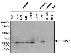
- Experimental details
- Western blot analysis of Heat Shock Protein 27 (HSP27) was performed by loading 50 µg of the indicated whole cell lysates per well onto a 4-20% Tris-HCl polyacrylamide gel. Proteins were transferred to a PVDF membrane and blocked with 5% BSA/TBST for at least 1 hour. The membrane was probed with a HSP27 monoclonal antibody (Product # MA3-015) at a dilution of 1:1000 overnight at 4°C on a rocking platform, washed in TBS-0.1%Tween 20, and probed with a goat anti-mouse IgG-HRP secondary antibody (Product # 31430) at a dilution of 1:20,000 for at least 1 hour. Chemiluminescent detection was performed using SuperSignal West Pico (Product # 34080).
- Submitted by
- Invitrogen Antibodies (provider)
- Main image
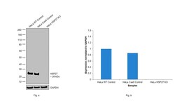
- Experimental details
- Knockout of HSP27 was achieved by CRISPR-Cas9 genome editing using LentiArray™ Lentiviral sgRNA (Product # A32042, Assay ID CRISPR990059_LV) and LentiArray Cas9 Lentivirus (Product # A32064). Western blot analysis of HSP27 was performed by loading 30 µg of HeLa Wild type (Lane 1), HeLa Cas9 (Lane 2) and HeLa HSP27 KO (Lane 3) whole cell extracts. The samples were electrophoresed using NuPAGE™ Novex™ 4-12% Bis-Tris Protein Gel (Product # NP0321BOX). Resolved proteins were then transferred onto a nitrocellulose membrane (Product # IB23001) by iBlot® 2 Dry Blotting System (Product # IB21001). The blot was probed with Anti-HSP27 Monoclonal Antibody (G3.1) (Product # MA3-015, 1:1,000 dilution) and Goat anti-Mouse IgG (H+L) Superclonal™ Recombinant Secondary Antibody, HRP (Product # A28177, 1:4,000 dilution) using the iBright FL 1000 (Product # A32752). Chemiluminescent detection was performed using Novex® ECL Chemiluminescent Substrate Reagent Kit (Product # WP20005). Loss of signal upon CRISPR mediated knockout (KO) using the LentiArray™ CRISPR product line confirms that antibody is specific to HSP27.
- Submitted by
- Invitrogen Antibodies (provider)
- Main image
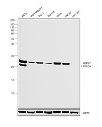
- Experimental details
- Western blot was performed using Anti-HSP27 Monoclonal Antibody (G3.1)(Product # MA3-015) and a 25kDa band corresponding to HSP27 was observed across cell lines tested except HT-1080 which is reported to be negative. Whole cell extracts (30 µg lysate) of MCF7 (Lane 1), MDA-MB-231 (Lane 2), PC-3 (Lane 3), DU 145 (Lane 4), HeLa (Lane 5), LNCaP (Lane 6) and HT-1080 (Lane 7) were electrophoresed using NuPAGE™ 12% Bis-Tris Protein Gel (Product # NP0342BOX). Resolved proteins were then transferred onto a Nitrocellulose membrane (Product # IB23001) by iBlot® 2 Dry Blotting System (Product # IB21001). The blot was probed with the primary antibody (1:1000 dilution) and detected by chemiluminescence with Goat anti-Mouse IgG (H+L) Superclonal™ Recombinant Secondary Antibody, HRP (Product # A28177,1:4000 dilution) using the iBright FL 1000 (Product # A32752). Chemiluminescent detection was performed using Novex® ECL Chemiluminescent Substrate Reagent Kit (Product # WP20005).
Supportive validation
- Submitted by
- Invitrogen Antibodies (provider)
- Main image
- Experimental details
- Immunofluorescent analysis of Heat Shock Protein 27 using Anti-Heat Shock Protein 27 Monoclonal Antibody (G3.1) (Product # MA3-015) shows staining in C6 Cells. Heat Shock Protein 27 staining (green), F-Actin staining with Phalloidin (red) and nuclei with DAPI (blue) is shown. Cells were grown on chamber slides and fixed with formaldehyde prior to staining. Cells were probed without (control) or with or an antibody recognizing Heat Shock Protein 27 (Product # MA3-015) at a dilution of 1:20 over night at 4°C, washed with PBS and incubated with a DyLight-488 conjugated secondary antibody (Product # 35503, Goat Anti-Mouse). Images were taken at 60X magnification.
- Submitted by
- Invitrogen Antibodies (provider)
- Main image
- Experimental details
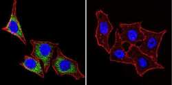
- Immunofluorescent analysis of Heat Shock Protein 27 using Anti-Heat Shock Protein 27 Monoclonal Antibody (G3.1) (Product # MA3-015) shows staining in Hela Cells. Heat Shock Protein 27 staining (green), F-Actin staining with Phalloidin (red) and nuclei with DAPI (blue) is shown. Cells were grown on chamber slides and fixed with formaldehyde prior to staining. Cells were probed without (control) or with or an antibody recognizing Heat Shock Protein 27 (Product # MA3-015) at a dilution of 1:200 over night at 4°C, washed with PBS and incubated with a DyLight-488 conjugated secondary antibody (Product # 35503, Goat Anti-Mouse). Images were taken at 60X magnification.
- Submitted by
- Invitrogen Antibodies (provider)
- Main image
- Experimental details
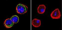
- Immunofluorescent analysis of Heat Shock Protein 27 using Anti-Heat Shock Protein 27 Monoclonal Antibody (G3.1) (Product # MA3-015) shows staining in U251 Cells. Heat Shock Protein 27 staining (green), F-Actin staining with Phalloidin (red) and nuclei with DAPI (blue) is shown. Cells were grown on chamber slides and fixed with formaldehyde prior to staining. Cells were probed without (control) or with or an antibody recognizing Heat Shock Protein 27 (Product # MA3-015) at a dilution of 1:200 over night at 4°C, washed with PBS and incubated with a DyLight-488 conjugated secondary antibody (Product # 35503, Goat Anti-Mouse). Images were taken at 60X magnification.
- Submitted by
- Invitrogen Antibodies (provider)
- Main image
- Experimental details
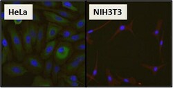
- Immunofluorescent analysis of Heat Shock Protein 27 (HSP27) (green) in HeLa cells and negative control NIH3T3 cells. Formalin fixed cells were permeabilized with 0.1% Triton X-100 in TBS for 10 minutes at room temperature and blocked with 1% Blocker BSA (Product # 37525) for 15 minutes at room temperature. Cells were probed with a HSP27 monoclonal antibody (Product # MA3-015), at a dilution of 1:50 for at least 1 hour at room temperature, washed with PBS, and incubated with DyLight 488 goat anti-mouse IgG secondary antibody (Product # 35502) at a dilution of 1:400 for 30 minutes at room temperature. F-Actin (red) was stained with DyLight 554 phalloidin (Product # 21834) and nuclei (blue) were stained with Hoechst 33342 dye (Product # 62249). Images were taken on a Thermo Scientific ArrayScan at 20X magnification.
- Submitted by
- Invitrogen Antibodies (provider)
- Main image
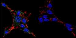
- Experimental details
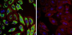
- Immunofluorescent analysis of Heat Shock Protein 27 (Hsp27, green) in HeLa cells. Formalin fixed cells were permeabilized with 0.1% Triton X-100 in TBS for 10 minutes at room temperature and blocked with 1% Blocker BSA (Product # 37525) for 15 minutes at room temperature. Cells were probed with (left panel) or without (right panel) a Hsp27 monoclonal antibody (Product # MA3-015), at a dilution of 1:100 for at least 1 hour at room temperature, washed with PBS, and incubated with DyLight 488 goat-anti-mouse IgG secondary antibody (Product # 35502) at a dilution of 1:400 for 30 minutes at room temperature. F-Actin (red) was stained with Dylight 554 phalloidin (Product # 21834), and nuclei (blue) were stained with Hoechst 33342 dye (Product # 62249). Images were taken on a Thermo Scientific ArrayScan or ToxInsight at 20X magnification.
Supportive validation
- Submitted by
- Invitrogen Antibodies (provider)
- Main image
- Experimental details
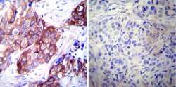
- Immunohistochemistry was performed on cancer biopsies of deparaffinized Human breast carcinoma tissues. To expose target proteins, heat induced antigen retrieval was performed using 10mM sodium citrate (pH6.0) buffer, microwaved for 8-15 minutes. Following antigen retrieval tissues were blocked in 3% BSA-PBS for 30 minutes at room temperature. Tissues were then probed at a dilution of 1:500 with a mouse monoclonal antibody recognizing Heat Shock Protein 27 (Product # MA3-015) or without primary antibody (negative control) overnight at 4°C in a humidified chamber. Tissues were washed extensively with PBST and endogenous peroxidase activity was quenched with a peroxidase suppressor. Detection was performed using a biotin-conjugated secondary antibody and SA-HRP, followed by colorimetric detection using DAB. Tissues were counterstained with hematoxylin and prepped for mounting.
- Submitted by
- Invitrogen Antibodies (provider)
- Main image
- Experimental details
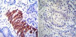
- Immunohistochemistry was performed on cancer biopsies of deparaffinized Human colon carcinoma tissues. To expose target proteins, heat induced antigen retrieval was performed using 10mM sodium citrate (pH6.0) buffer, microwaved for 8-15 minutes. Following antigen retrieval tissues were blocked in 3% BSA-PBS for 30 minutes at room temperature. Tissues were then probed at a dilution of 1:200 with a mouse monoclonal antibody recognizing Heat Shock Protein 27 (Product # MA3-015) or without primary antibody (negative control) overnight at 4°C in a humidified chamber. Tissues were washed extensively with PBST and endogenous peroxidase activity was quenched with a peroxidase suppressor. Detection was performed using a biotin-conjugated secondary antibody and SA-HRP, followed by colorimetric detection using DAB. Tissues were counterstained with hematoxylin and prepped for mounting.
- Submitted by
- Invitrogen Antibodies (provider)
- Main image
- Experimental details
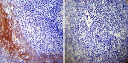
- Immunohistochemistry was performed on normal deparaffinized Human tonsil tissue tissues. To expose target proteins, heat induced antigen retrieval was performed using 10mM sodium citrate (pH6.0) buffer, microwaved for 8-15 minutes. Following antigen retrieval tissues were blocked in 3% BSA-PBS for 30 minutes at room temperature. Tissues were then probed at a dilution of 1:500 with a mouse monoclonal antibody recognizing Heat Shock Protein 27 (Product # MA3-015) or without primary antibody (negative control) overnight at 4°C in a humidified chamber. Tissues were washed extensively with PBST and endogenous peroxidase activity was quenched with a peroxidase suppressor. Detection was performed using a biotin-conjugated secondary antibody and SA-HRP, followed by colorimetric detection using DAB. Tissues were counterstained with hematoxylin and prepped for mounting.
Supportive validation
- Submitted by
- Invitrogen Antibodies (provider)
- Main image
- Experimental details
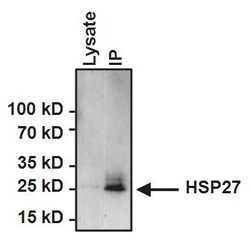
- Immunoprecipitation of Heat Shock Protein 27 (HSP27) was performed on HeLa cells. Antigen:antibody complexes were formed by incubating 500 µg whole cell lysate with 2 µg of HSP27 monoclonal antibody (Product # MA3-015) overnight on a rocking platform at 4°C. The immune complexes were captured on 50 µL Protein A/G Plus Agarose (Product # 20423), washed extensively, and eluted with 5X Lane Marker Reducing Sample Buffer (Product # 39000). HeLa cell lysate (25 µg) was loaded as a positive control. Samples were resolved on a 4-20% Tris-HCl polyacrylamide gel, transferred to a PVDF membrane, and blocked with 5% BSA/TBST for at least 1 hour. The membrane was probed with a HSP27 monoclonal antibody (Product # MA3-015) at a dilution of 1:1000 overnight rotating at 4°C, washed in TBST, and probed with Clean-Blot IP Detection Reagent-HRP (Product # 21230) at a dilution of 1:1000 for at least one hour. Chemiluminescent detection was performed using SuperSignal West Dura (Product # 34075).
- Submitted by
- Invitrogen Antibodies (provider)
- Main image
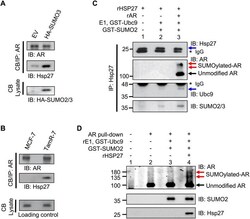
- Experimental details
- Fig. 3 AR interacts with and is a substrate for the SUMO E3 ligase Hsp27. a Ectopic induction of a hyperSUMO environment in MCF-7 cells with HA-SUMO3 increases the interaction between AR and Hsp27. b AR-Hsp27 interaction complex is enhanced endogenously in the naturally occurring hyperSUMO environment of TamR-7 cells. a - b Interactions with chromatin-bound (CB) AR were analyzed by western blot. c Immunoprecipitation of Hsp27 was performed to assess its binding with AR, Ubc9 and SUMO-2/3 in a cell-free system. d SUMOylation of AR is enhanced under in vitro conditions when recombinant Hsp27 is added. Details are found in (Additional file 1 )
 Explore
 Validate
 Learn
Learn