Explore
 Validate
 Learn
Learn Western blot
 Immunohistochemistry
Antibody data
- Antibody Data
- Antigen structure
- References [1]
- Comments [0]
- Validations
- Immunohistochemistry [3]
- Other assay [4]
Submit
Validation data
Reference
Comment
Report error
- Product number
- PA5-50759 - Provider product page

- Provider
- Invitrogen Antibodies
- Product name
- SIGLEC15 Polyclonal Antibody
- Antibody type
- Polyclonal
- Antigen
- Synthetic peptide
- Description
- The antibody detects endogenous levels of total SIGLEC15 protein.
- Reactivity
- Human, Mouse
- Host
- Rabbit
- Isotype
- IgG
- Vial size
- 100 µL
- Concentration
- 0.9 mg/mL
- Storage
- -20°C
Submitted references LINC00973 is involved in cancer immune suppression through positive regulation of Siglec-15 in clear-cell renal cell carcinoma.
Liu Y, Li X, Zhang C, Zhang H, Huang Y
Cancer science 2020 Oct;111(10):3693-3704
Cancer science 2020 Oct;111(10):3693-3704
No comments: Submit comment
Supportive validation
- Submitted by
- Invitrogen Antibodies (provider)
- Main image
- Experimental details
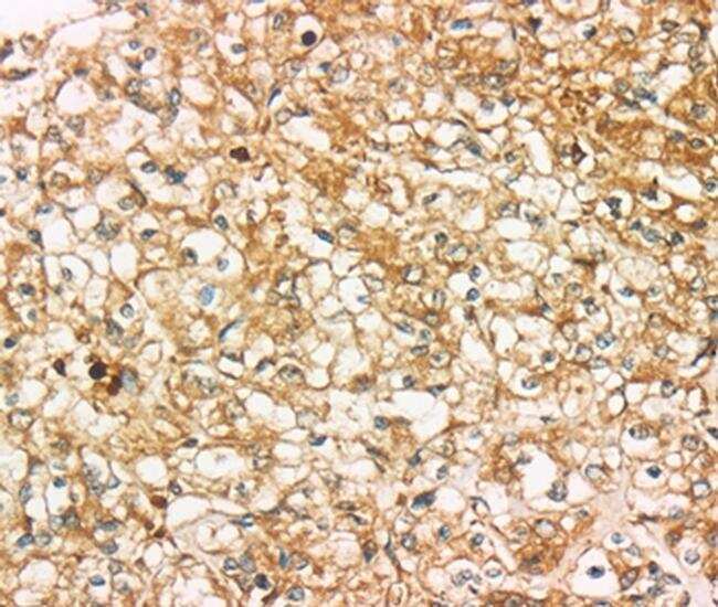
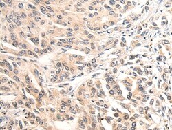
- Immunohistochemical analysis of SIGLEC15 in paraffin-embedded Human prostate cancer tissue. Samples were probed using a SIGLEC15 polyclonal antibody (Product # PA5-50759) at a dilution of 1/35.
- Submitted by
- Invitrogen Antibodies (provider)
- Main image
- Experimental details
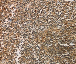
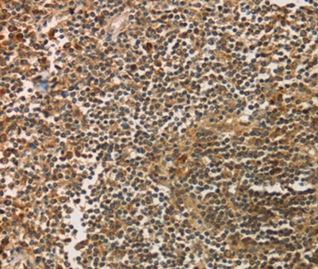
- Immunohistochemical analysis of SIGLEC15 in paraffin-embedded Human tonsil cancer tissue. Samples were probed using a SIGLEC15 polyclonal antibody (Product # PA5-50759) at a dilution of 1/35.
- Submitted by
- Invitrogen Antibodies (provider)
- Main image
- Experimental details
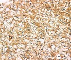
- Immunohistochemical analysis of SIGLEC15 inparaffin-embedded Human liver cancer tissue using SIGLEC15 Polyclonal Antibody (Product # PA5-50759) at a 1:50 dilution. (Original magnification: x200).

Supportive validation
- Submitted by
- Invitrogen Antibodies (provider)
- Main image
- Experimental details
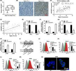
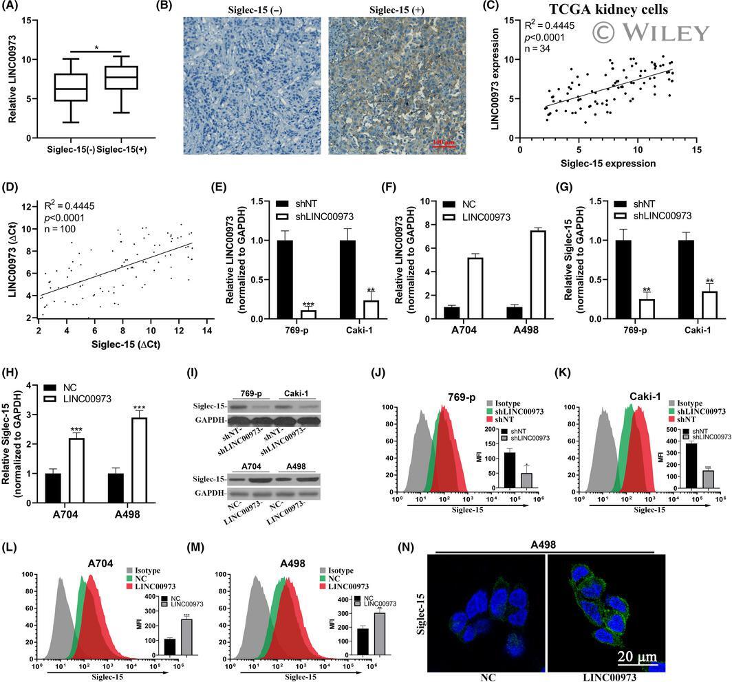
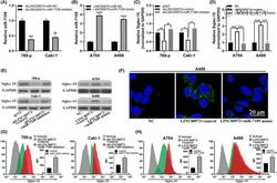
- 2 FIGURE LINC00973 upregulated Siglec-15 in clear-cell renal cell carcinoma (ccRCC) cells. (A) Comparison of LINC00973 abundance in Siglec-15 negative and positive ccRCC tumors. (B) Representative images of Siglec-15 immunohistochemistry (IHC) staining results. (C) Correlation analysis of endogenous LINC00973 and Siglec-15 expression in publicly available TCGA kidney cells (n = 34). (D) Correlation analysis of LINC00973 and Siglec-15 in clinical ccRCC tumor samples (n = 100). (E) Establishment of LINC00973-knockdown cell lines in 769-p and Caki-1 cells was validated by real-time PCR. (F) Establishment of LINC00973-overexpressing cell lines in A704 and A498 cells was validated by real-time PCR. (G) Relative expression of Siglec-15 transcripts in LINC00973-deficient 769-p and Caki-1 cells. (H). Relative expression of Siglec-15 transcripts in LINC00973-proficient A704 and A498 cells. (I) Western blot analysis of Siglec-15 protein in LINC00973 knockdown or overexpressing cells. Flow cytometry analysis of cell surface Siglec-15 abundance in response to LINC00973 knockdown in 769-p (J) and Caki-1 (K) cells, and LINC00973-overexpressing in A704 (L) and A498 (M). N. Immunofluorescence staining of cell surface Siglec-15 of A498 in response to either control (NC) or LINC00973 overexpression
- Submitted by
- Invitrogen Antibodies (provider)
- Main image
- Experimental details
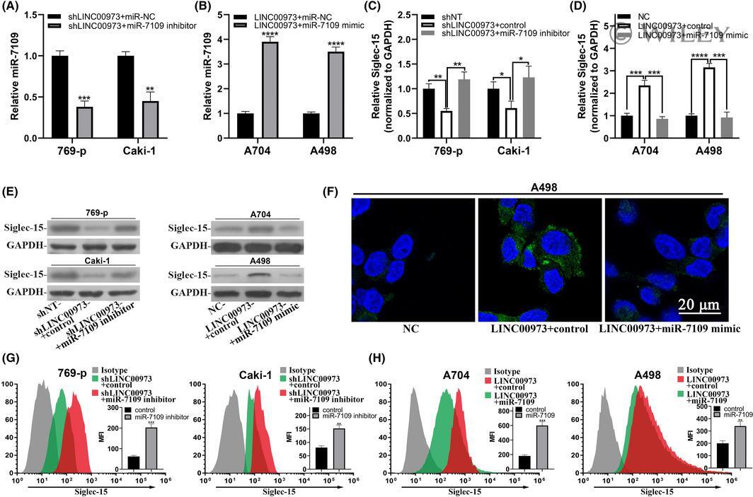
- 3 FIGURE LINC00973-upregulated Siglec-15 was dependent on miR-7109. (A) Real-time PCR analysis of endogenous miR-7109 in LINC00973-deficient 769-p and Caki-1 cells in response to either negative control or miR-7109 inhibitor. (B) Real-time PCR analysis of endogenous miR-7109 in LINC00973-proficient A704 and A498 cells in response to either control or miR-7901 mimic. (C) Quantitative analysis of Siglec-15 transcript in LINC00973 intact or depleted 769-p and Caki-1 cells treated with control or miR-7109 inhibitor. (D) Quantitative analysis of Siglec-15 transcript in LINC00973 naive or overexpressing A704 and A498 cells treated with control or miR-7109 mimics. (E) Western blot results of Siglec-15 protein in the cells as described in C and D. (F) Immunofluorescence staining of cell surface Siglec-15 in A498 cells expressing either naive or ectopic LINC00973, and transfected with either control or miR-7109 mimics. (G, H) Flow cytometry analysis of cell surface abundance of Siglec-15 in cells as specified in panel E
- Submitted by
- Invitrogen Antibodies (provider)
- Main image

- Experimental details
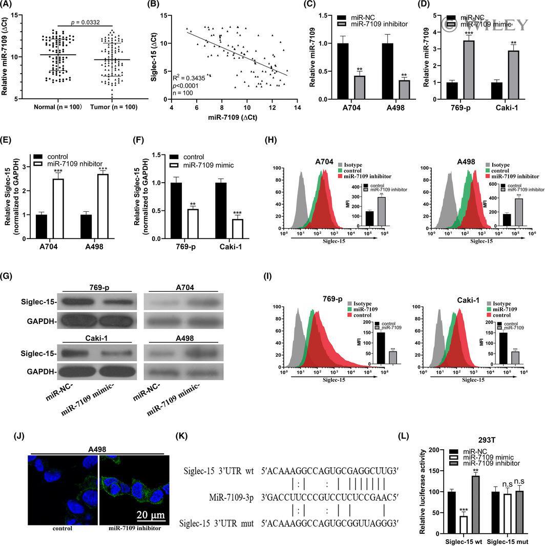
- 4 FIGURE Siglec-15 was a direct target of miR-7109 in clear-cell renal cell carcinoma (ccRCC). (A) Real-time PCR analysis of miR-7109 expression in ccRCC tumor samples with paired adjacent normal tissues. (B) Correlation analysis between miR-7109 and Siglec-15 in ccRCC tissue samples (n = 100). (C) Change of miR-7109 expression in both A704 and A498 cells in response to miR-7109 inhibitor. (D) Change of miR-7109 expression in both 769-p and Caki-1 cells in response to miR-7109 mimic. (E) Endogenous Siglec-15 transcripts were quantified in both A704 and A498 cells transfected with either control or miR-7109 inhibitor. (F) Endogenous Siglec-15 transcripts were quantified in both 769-p and Caki-1 cells transfected with either control or miR-7109 mimics. (G) Western blot analysis of Siglec-15 proteins in the same cells as in panel E and F. (H) Flow cytometry analysis of cell surface Siglec-15 abundance in A704 and A498 cells in response to miR-7109 inhibitor. (I) Flow cytometry analysis of cell surface Siglec-15 abundance in 769-p and Caki-1 cells in response to miR-7109 mimics. (J) Immunofluorescence staining of cell surface Siglec-15 in A498 cells transfected with either control or miR-7109 inhibitor. (K) Alignment between miR-7109 seed region and both wild-type and putative binding site-mutated Siglec-15 3'UTR. (L) Luciferase reporter analysis of the potential regulatory effects of miR-7109 on Siglec-15 in 293T cells, which were transfected with control, miR-7109 mimics, and m
- Submitted by
- Invitrogen Antibodies (provider)
- Main image

- Experimental details
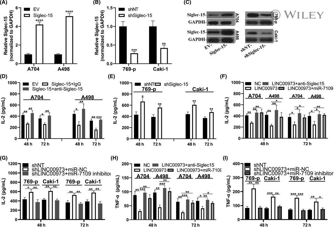
- 6 FIGURE LINC00973-miR-7109-Siglec-15 axis involved in immune activations in clear-cell renal cell carcinoma (ccRCC) cells. (A) Establishment of Siglec-15 overexpression cells in A704 and A498 was confirmed by real-time PCR. (B) Establishment of Siglec-15 knockdown cells in 769-p and Caki-1 cells was confirmed by real-time PCR. (C) Western blot results of relative expression of Siglec-15 protein in cells described in (A) and (B). (D) Interleukin-2 (IL-2) production analysis in Jurkat: A704/A498 (EV or Siglec-15, 2:1) co-culture system. IL-2 was quantified with an ELISA kit at 48 and 72 h, respectively. (E) IL-2 production analysis in Jurkat: 769-p/Caki-1 (shNT or shSiglec-15, 2:1) co-culture system. (F) IL-2 production analysis of Jurkat co-culture system with both A704 and A498 cells transfected with control or LINC00973 in combination with either miR-NC or miR7109 mimics. (G) IL-2 production analysis of Jurkat co-culture system with both 769-p and Caki-1 cells transfected with negative control or shLINC0973 in combination with either miR-NC or miR-7109 inhibitor. (H) Tumor necrosis factor-alpha (TNF-alpha) secretion analysis of Jurkat co-culture system with both A704 and A498 cells transfected with control or LINC00973 in combination with either miR-NC or miR-7109 mimics. (I) TNF-alpha secretion analysis of Jurkat co-culture system with both 769-p and Caki-1 cells transfected with either negative control or shLINC00973 in combination with either miR-NC or miR-7109 inhibitor