Explore
 Validate
 Learn
Learn Immunocytochemistry
Antibody data
- Antibody Data
- Antigen structure
- References [1]
- Comments [0]
- Validations
- Immunocytochemistry [1]
- Immunohistochemistry [3]
- Other assay [1]
Submit
Validation data
Reference
Comment
Report error
- Product number
- PA5-63228 - Provider product page

- Provider
- Invitrogen Antibodies
- Product name
- CASKIN1 Polyclonal Antibody
- Antibody type
- Polyclonal
- Antigen
- Recombinant full-length protein
- Reactivity
- Human
- Host
- Rabbit
- Isotype
- IgG
- Vial size
- 100 µL
- Concentration
- 0.2 mg/mL
- Storage
- Store at 4°C short term. For long term storage, store at -20°C, avoiding freeze/thaw cycles.
Submitted references The Human Hippocampus in Parkinson's Disease: An Integrative Stereological and Proteomic Study.
Villar-Conde S, Astillero-Lopez V, Gonzalez-Rodriguez M, Villanueva-Anguita P, Saiz-Sanchez D, Martinez-Marcos A, Flores-Cuadrado A, Ubeda-Bañon I
Journal of Parkinson's disease 2021;11(3):1345-1365
Journal of Parkinson's disease 2021;11(3):1345-1365
No comments: Submit comment
Supportive validation
- Submitted by
- Invitrogen Antibodies (provider)
- Main image
- Experimental details
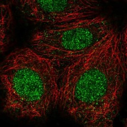
- Immunofluorescent staining of CASKIN1 in human cell line MCF7 using a CASKIN1 Polyclonal Antibody (Product # PA5-63228) shows localization to nucleus.

Supportive validation
- Submitted by
- Invitrogen Antibodies (provider)
- Main image
- Experimental details
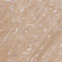
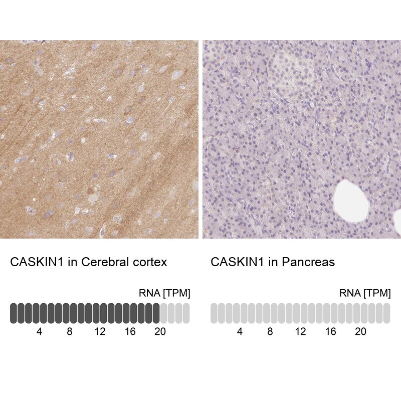
- Immunohistochemical staining of CASKIN1 in human cerebral cortex tissue shows moderate cytoplasmic positivity in subset of neuronal cells and neuropil. Samples were probed using a CASKIN1 Polyclonal Antibody (Product # PA5-63228).
- Submitted by
- Invitrogen Antibodies (provider)
- Main image
- Experimental details
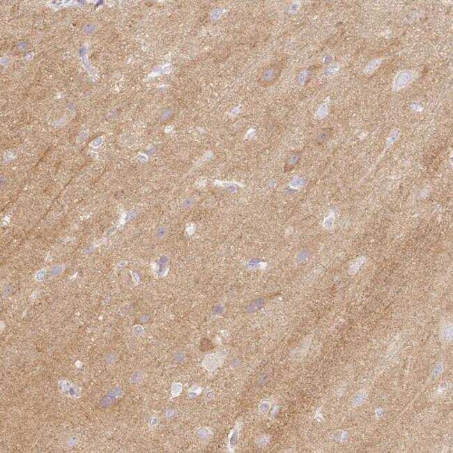
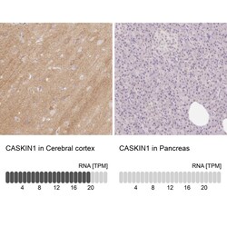
- Immunohistochemical staining of CASKIN1 in human cerebral cortex and pancreas tissues using CASKIN1 Polyclonal Antibody (Product # PA5-63228). Corresponding CASKIN1 RNA-seq data are presented for the same tissues.
- Submitted by
- Invitrogen Antibodies (provider)
- Main image
- Experimental details
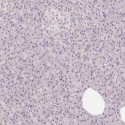
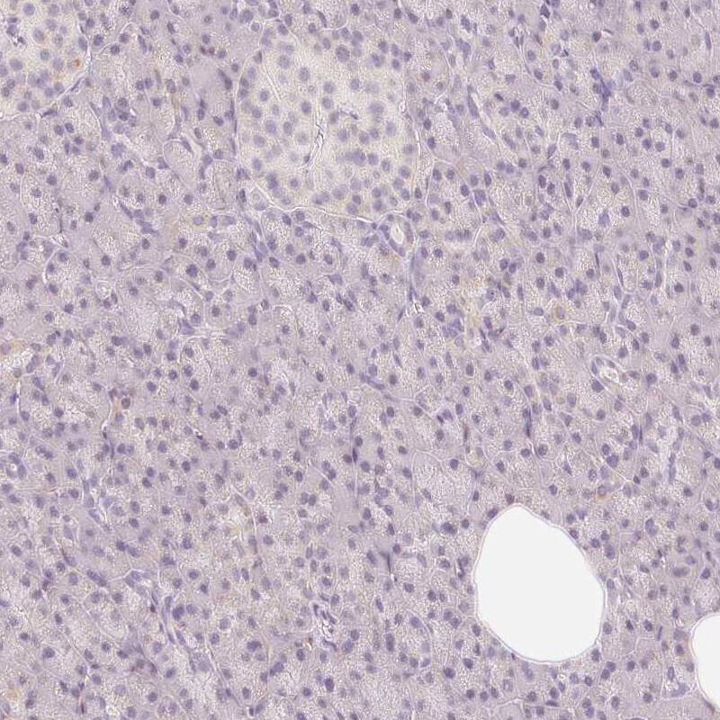
- Immunohistochemical staining of CASKIN1 in human pancreas using CASKIN1 Polyclonal Antibody (Product # PA5-63228) shows low expression as expected.
Supportive validation
- Submitted by
- Invitrogen Antibodies (provider)
- Main image
- Experimental details
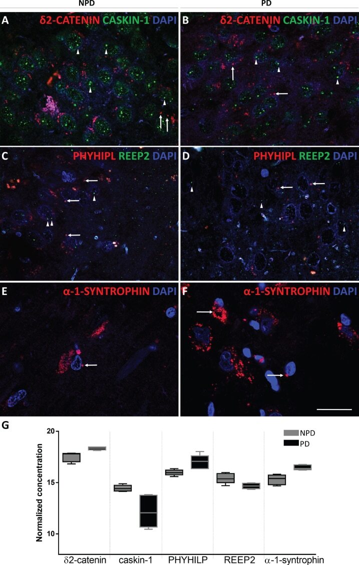
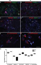
- Fig. 8 Coronal sections of the human hippocampus immunofluorescently stained for delta2-catenin (RED) and caskin-1 (GREEN) in NPD (A) and PD (B), REEP2 (GREEN) and PHYHIPL (RED) in NPD (C) and PD (D) and alpha-syntrophin (RED) in NPD (E) and NPD (F). Arrow heads and arrows point green and red markers, respectively. Scale bar 20mum (A-F). The mean+-SD of selected proteins comparing NPD and PD (G).