Explore
 Validate
 Learn
Learn Western blot
Antibody data
- Antibody Data
- Antigen structure
- References [1]
- Comments [0]
- Validations
- Western blot [1]
- Immunocytochemistry [1]
- Immunohistochemistry [6]
Submit
Validation data
Reference
Comment
Report error
- Product number
- AMAb90501 - Provider product page

- Provider
- Atlas Antibodies
- Proper citation
- Atlas Antibodies Cat#AMAb90501, RRID:AB_2665567
- Product name
- Anti-SOX11
- Antibody type
- Monoclonal
- Reactivity
- Human, Mouse
- Host
- Mouse
- Conjugate
- Unconjugated
- Antigen sequence
FMVWSKIERRKIMEQSPDMHNAEISKRLGKRWKML
KDSEKIPFIREAERLRLKHMADYPDYKYRPRKKPK
MDPSAKPSASQSPEKSAAGGGGGSAGGGAGGAKTS
KGSSKK- Epitope
- Binds to an epitope located within the peptide sequence IPFIREAERL as determined by overlapping synthetic peptides.
- Isotype
- IgG
- Antibody clone number
- CL0142
- Vial size
- 100 µl
- Storage
- Store at +4°C for short term storage. Long time storage is recommended at -20°C.
Submitted references Assessment of SOX11 expression in routine lymphoma tissue sections: characterization of new monoclonal antibodies for diagnosis of mantle cell lymphoma.
Soldini D, Valera A, Solé C, Palomero J, Amador V, Martin-Subero JI, Ribera-Cortada I, Royo C, Salaverria I, Beà S, Gonzalvo E, Johannesson H, Herrera M, Colomo L, Martinez A, Campo E
The American journal of surgical pathology 2014 Jan;38(1):86-93
The American journal of surgical pathology 2014 Jan;38(1):86-93
No comments: Submit comment
Supportive validation
- Submitted by
- Atlas Antibodies (provider)
- Main image
- Experimental details
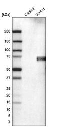
- Western blot analysis in control (vector only transfected HEK293T lysate) and sOX11 over-expression lysate (Co-expressed with a C-terminal myc-DDK tag (~3.1 kDa) in mammalian HEK293T cells, LY418895).

Supportive validation
- Submitted by
- Atlas Antibodies (provider)
- Main image
- Experimental details
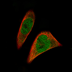
- Immunofluorescence staining of RH-30 cells using the Anti-SOX11 monoclonal antibody, showing specific staining in the nucleus and cytosol in green. Microtubule- and nuclear probes are visualized in red and blue, respectively (where available).
- Sample type
- HUMAN

Supportive validation
- Submitted by
- Atlas Antibodies (provider)
- Main image
- Experimental details
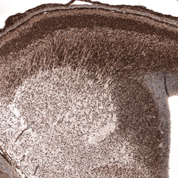
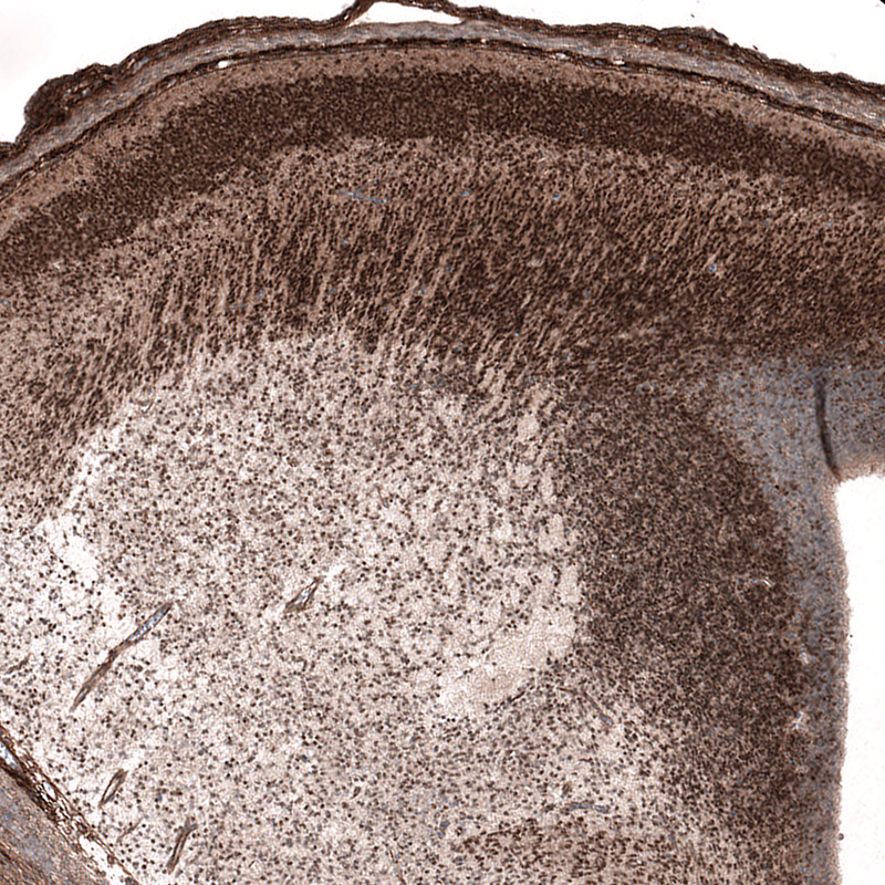
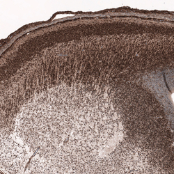
- Immunohistochemical staining of mouse embryo E14 shows nuclear immunoreactivity in the developing forebrain neurons.
- Submitted by
- Atlas Antibodies (provider)
- Main image
- Experimental details
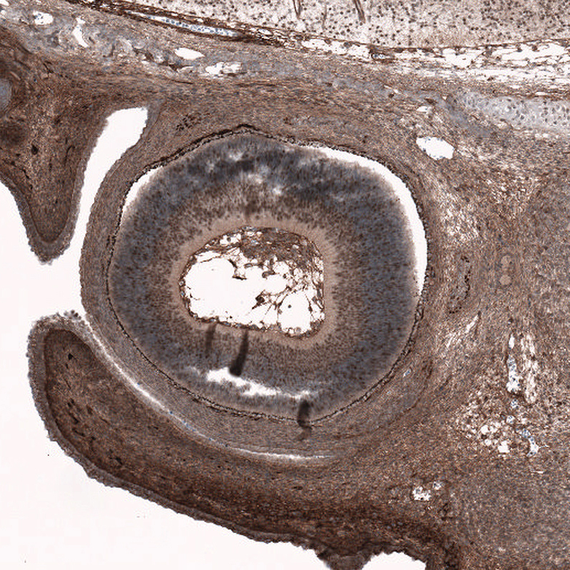
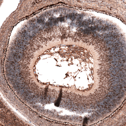
- Immunohistochemical staining of mouse embryo E14 shows nuclear immunoreactivity in the developing eye.

- Submitted by
- Atlas Antibodies (provider)
- Main image
- Experimental details
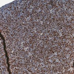
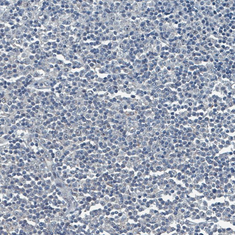
- Immunohistochemical staining of human mantle cell lymphoma shows moderate to strong nuclear positivity in tumor cells.
- Submitted by
- Atlas Antibodies (provider)
- Main image
- Experimental details
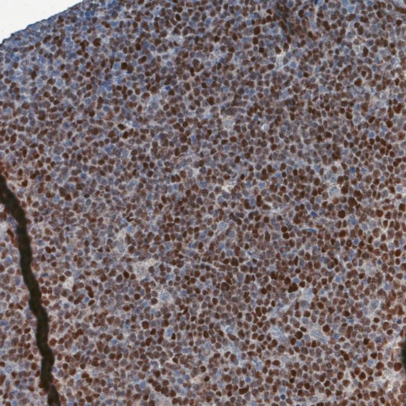
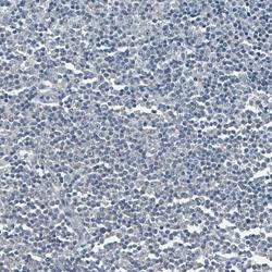
- Immunohistochemical staining of human chronic lymphocytic leukemia shows no nuclear positivity in tumor cells as expected.
- Submitted by
- Atlas Antibodies (provider)
- Main image
- Experimental details
- Immunohistochemical staining of mouse embryo E14 shows strong nuclear positivity in the developing forebrain.

- Submitted by
- Atlas Antibodies (provider)
- Main image

- Experimental details
- Immunohistochemical staining of eye in mouse embryo E14 retina shows moderate nuclear positivity in developing retina.