Explore
 Validate
 Learn
Learn701225
antibody from Invitrogen Antibodies
Targeting: CXCL10
C7, crg-2, gIP-10, IFI10, INP10, IP-10, mob-1, SCYB10
 Western blot
Antibody data
- Antibody Data
- Antigen structure
- References [2]
- Comments [0]
- Validations
- Western blot [1]
- Immunocytochemistry [1]
- Immunohistochemistry [1]
Submit
Validation data
Reference
Comment
Report error
- Product number
- 701225 - Provider product page

- Provider
- Invitrogen Antibodies
- Product name
- CXCL10 Recombinant Rabbit Monoclonal Antibody (10H11L3)
- Antibody type
- Monoclonal
- Antigen
- Other
- Reactivity
- Human, Mouse
- Host
- Rabbit
- Isotype
- IgG
- Antibody clone number
- 10H11L3
- Vial size
- 100 µg
- Concentration
- 0.5 mg/mL
- Storage
- Store at 4°C short term. For long term storage, store at -20°C, avoiding freeze/thaw cycles.
Submitted references Sensory neuron dysfunction in orthotopic mouse models of colon cancer.
Immune synapses between mast cells and γδ T cells limit viral infection.
Balogh M, Zhang J, Gaffney CM, Kalakuntla N, Nguyen NT, Trinh RT, Aguilar C, Pham HV, Milutinovic B, Nichols JM, Mahalingam R, Shepherd AJ
Journal of neuroinflammation 2022 Aug 12;19(1):204
Journal of neuroinflammation 2022 Aug 12;19(1):204
Immune synapses between mast cells and γδ T cells limit viral infection.
Mantri CK, St John AL
The Journal of clinical investigation 2019 Mar 1;129(3):1094-1108
The Journal of clinical investigation 2019 Mar 1;129(3):1094-1108
No comments: Submit comment
Supportive validation
- Submitted by
- Invitrogen Antibodies (provider)
- Main image
- Experimental details
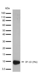
- Western blot analysis of mouse IP-10 recombinant protein using an IP-10 recombinant rabbit monoclonal antibody (Product # 701225) at a dilution of 1 µg/mL. Samples were detected using chemiluminescence (ECL). Results show a band at ~8.9kDa.

Supportive validation
- Submitted by
- Invitrogen Antibodies (provider)
- Main image
- Experimental details
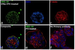
- Immunofluorescence analysis of C-X-C motif chemokine 10 was performed using 70% confluent log phase HT-29 cells and HT-29 cells treated with 50 ng/mL IFN gamma for 14 hr followed by 1x PTI treatment for 4h. The cells were fixed with 4% paraformaldehyde for 10 minutes, permeabilized with 0.1% Triton™ X-100 for 15 minutes, and blocked with 2% BSA for 45 minutes at room temperature. The cells were labeled with CXCL10 Recombinant Rabbit Monoclonal Antibody (10H11L3) (Product # 701225) at 1:200 in 0.1% BSA, incubated at 4 degree celsius overnight and then labeled with Donkey anti-Rabbit IgG (H+L) Highly Cross-Adsorbed Secondary Antibody, Alexa Fluor Plus 488 (Product # A32790), (1:2500), for 45 minutes at room temperature (Panel a: Green). Nuclei (Panel b:Blue) were stained with ProLong™ Diamond Antifade Mountant with DAPI (Product # P36962). F-actin (Panel c: Red) was stained with Rhodamine Phalloidin (Product # R415, 1:300). Panel d represents the merged image showing Cytosolic localization. Panel f represents control cells with no primary antibody to assess background. The images were captured at 60X magnification.

Supportive validation
- Submitted by
- Invitrogen Antibodies (provider)
- Main image
- Experimental details
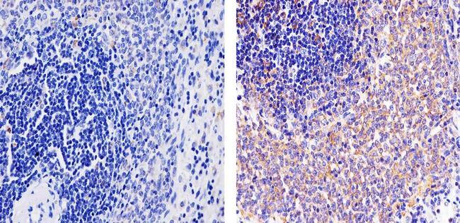
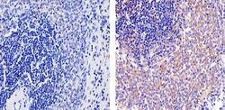
- Immunohistochemistry analysis of IP-10 showing staining in the cytoplasm of paraffin-embedded mouse spleen tissue (right) compared to a negative control without primary antibody (left). To expose target proteins, antigen retrieval was performed using 10mM sodium citrate (pH 6.0), microwaved for 8-15 min. Following antigen retrieval, tissues were blocked in 3% H2O2-methanol for 15 min at room temperature, washed with ddH2O and PBS, and then probed with a IP-10 (MS) Recombinant Rabbit Monoclonal Antibody (Product # 701225) diluted in 3% BSA-PBS at a dilution of 1:20 for 1 hour at 37ºC in a humidified chamber. Tissues were washed extensively in PBST and detection was performed using an HRP-conjugated secondary antibody followed by colorimetric detection using a DAB kit. Tissues were counterstained with hematoxylin and dehydrated with ethanol and xylene to prep for mounting.