 Explore
 Validate
 Learn
Learn Western blot
 Immunohistochemistry
Antibody data
- Antibody Data
- Antigen structure
- References [2]
- Comments [0]
- Validations
- Western blot [1]
- Immunohistochemistry [12]
Submit
Validation data
Reference
Comment
Report error
- Product number
- HPA001252 - Provider product page

- Provider
- Atlas Antibodies
- Proper citation
- Atlas Antibodies Cat#HPA001252, RRID:AB_1078906
- Product name
- Anti-FOXO1
- Antibody type
- Polyclonal
- Reactivity
- Human, Mouse
- Host
- Rabbit
- Conjugate
- Unconjugated
- Antigen sequence
LTSDSPPHNDIMTPVDPGVAQPNSRVLGQNVMMGP
NSVMSTYGSQASHNKMMNPSSHTHPGHAQQTSAVN
GRPLPHTVSTMPHTSGMNRLTQVKTPVQVPLPHPM
QMSALGGYSSVSSCNGYGRMGLLHQEKLPSDLDGM
FIERLDCDM- Isotype
- IgG
- Vial size
- 100 µl
- Storage
- Store at +4°C for short term storage. Long time storage is recommended at -20°C.
Submitted references Dual role of FoxA1 in androgen receptor binding to chromatin, androgen signalling and prostate cancer.
Variance decomposition of protein profiles from antibody arrays using a longitudinal twin model.
Sahu B, Laakso M, Ovaska K, Mirtti T, Lundin J, Rannikko A, Sankila A, Turunen JP, Lundin M, Konsti J, Vesterinen T, Nordling S, Kallioniemi O, Hautaniemi S, Jänne OA
The EMBO journal 2011 Sep 13;30(19):3962-76
The EMBO journal 2011 Sep 13;30(19):3962-76
Variance decomposition of protein profiles from antibody arrays using a longitudinal twin model.
Kato BS, Nicholson G, Neiman M, Rantalainen M, Holmes CC, Barrett A, Uhlén M, Nilsson P, Spector TD, Schwenk JM
Proteome science 2011 Nov 17;9:73
Proteome science 2011 Nov 17;9:73
No comments: Submit comment
Enhanced validation
- Submitted by
- Atlas Antibodies (provider)
- Enhanced method
- Recombinant expression validation
- Main image
- Experimental details
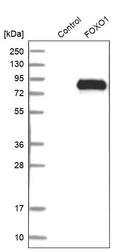
- Western blot analysis in control (vector only transfected HEK293T lysate) and FOXO1 over-expression lysate (Co-expressed with a C-terminal myc-DDK tag (~3.1 kDa) in mammalian HEK293T cells, LY419586).

Supportive validation
- Submitted by
- Atlas Antibodies (provider)
- Main image
- Experimental details
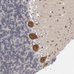
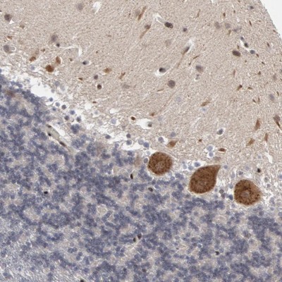
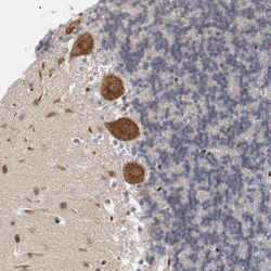
- Immunohistochemical staining of human cerebellum shows strong cytoplasmic positivity in Purkinje cells.
- Submitted by
- Atlas Antibodies (provider)
- Main image
- Experimental details
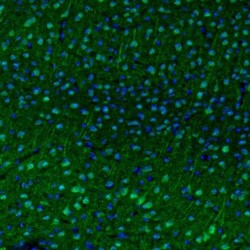
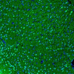
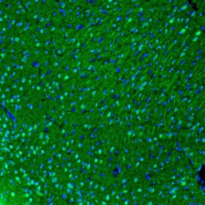
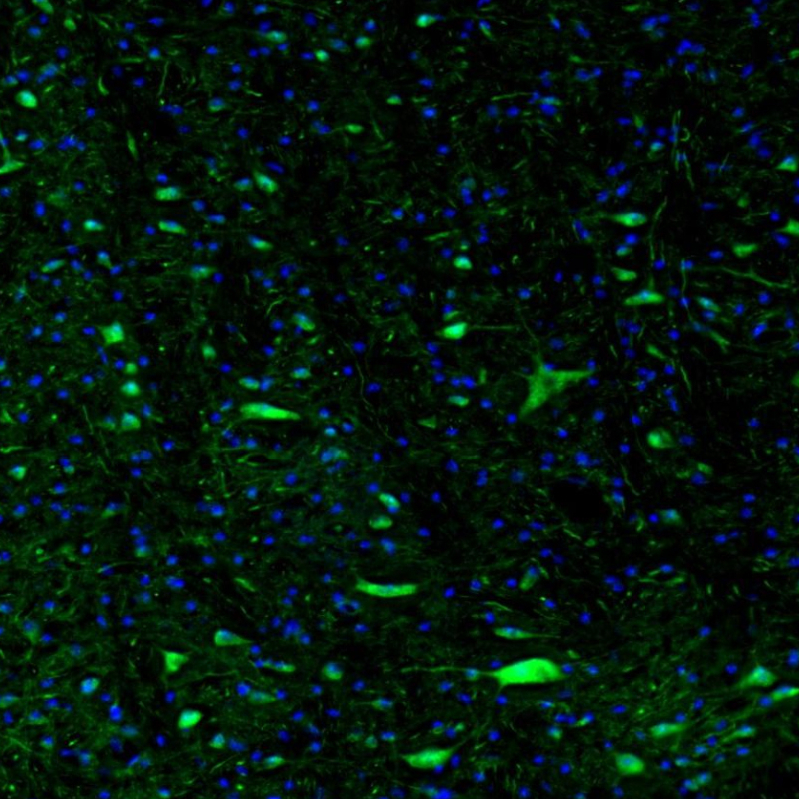
- Immunofluorescence staining of mouse visual cortex shows immunoreactivity in a subset of neurons.

- Submitted by
- Atlas Antibodies (provider)
- Main image

- Experimental details
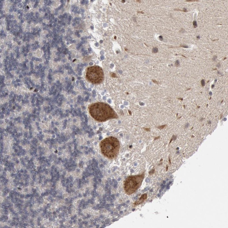
- Immunohistochemical staining of human cerebellum shows cytoplasmic positivity in Purkinje cells.
- Submitted by
- Atlas Antibodies (provider)
- Main image
- Experimental details
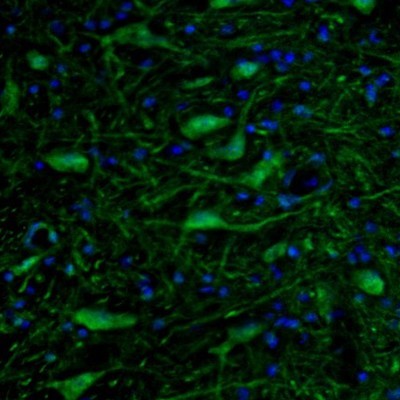
- Immunofluorescence staining of mouse insular cortex shows immunoreactivity in neuronal cells.
- Submitted by
- Atlas Antibodies (provider)
- Main image
- Experimental details
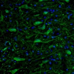
- Immunofluorescence staining of mouse pons shows immunoreactivity in neuronal cell bodies and processes in the motor trigeminal nucleus.
- Submitted by
- Atlas Antibodies (provider)
- Main image
- Experimental details
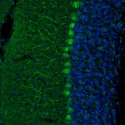
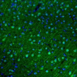
- Immunofluorescence staining of mouse cerebellum shows immunoreactivity in Purkinje cells.

- Submitted by
- Atlas Antibodies (provider)
- Main image

- Experimental details
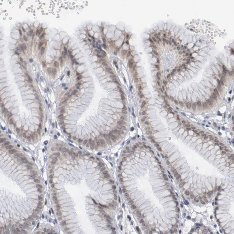
- Immunohistochemical staining of human stomach shows weak to moderate nuclear positivity in glandular cells.
- Sample type
- HUMAN
- Submitted by
- Atlas Antibodies (provider)
- Main image

- Experimental details
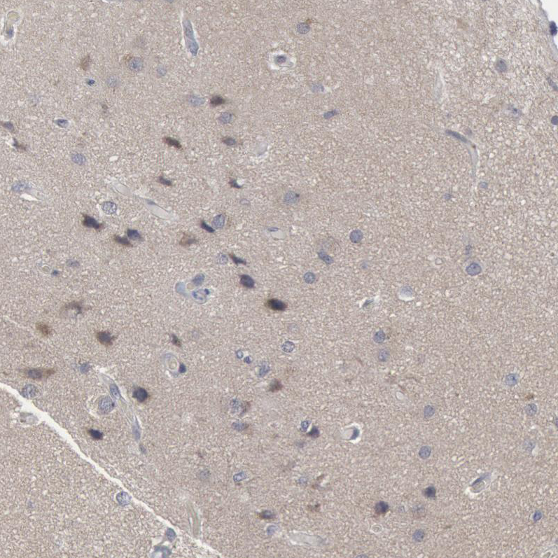
- Immunohistochemical staining of human cerebral cortex shows moderate to strong cytoplasmic positivity in neurons.
- Sample type
- HUMAN
- Submitted by
- Atlas Antibodies (provider)
- Main image
- Experimental details
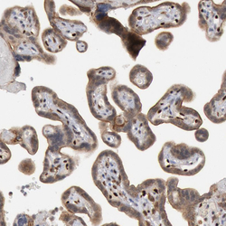
- Immunohistochemical staining of human placenta shows weak to moderate cytoplasmic positivity in trophoblastic cells.
- Sample type
- HUMAN

- Submitted by
- Atlas Antibodies (provider)
- Main image
- Experimental details
- Immunohistochemical staining of human cerebellum shows moderate to strong cytoplasmic positivity in purkinje cells.
- Sample type
- HUMAN

- Submitted by
- Atlas Antibodies (provider)
- Main image

- Experimental details
- Immunofluorescence staining of mouse brain shows moderate to strong positivity in more than 75 % of neurons in the pons
- Sample type
- MOUSE
- Submitted by
- Atlas Antibodies (provider)
- Main image
- Experimental details
- Immunofluorescence staining of mouse cerebral cortex shows moderate to strong positivity in more than 75 % of neurons in the visual cortex.
- Sample type
- MOUSE
