Explore
 Validate
 Learn
LearnAP09260PU-N
antibody from Acris Antibodies GmbH
Targeting: GLI3
ACLS, GCPS, PAP-A, PAPA, PAPA1, PAPB, PHS, PPDIV
 Western blot
 ELISA
ELISAAntibody data
- Antibody Data
- Antigen structure
- References [0]
- Comments [0]
- Validations
- Western blot [1]
- Immunohistochemistry [1]
Submit
Validation data
Reference
Comment
Report error
- Product number
- AP09260PU-N - Provider product page

- Provider
- Acris Antibodies GmbH
- Proper citation
- Acris Antibodies GmbH Cat#AP09260PU-N, RRID:AB_2035561
- Product name
- anti GLI3 (41-57)
- Antibody type
- Polyclonal
- Antigen
- Synthetic peptide corresponding to amino acids 41-57 of Human Gli-3 protein
- Reactivity
- Human, Mouse, Rat, Canine, Chicken/Avian, Simian, Xenopus
- Host
- Rabbit
- Isotype
- IgG
- Vial size
- 0.1 mg
- Concentration
- 1.0 mg/ml (by UV absorbance at 280 nm)
No comments: Submit comment
Supportive validation
- Submitted by
- Acris Antibodies GmbH (provider)
- Main image
- Experimental details
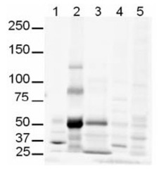
- Western blot using AP09260PU-N Gli-3 antibody shows detection of multiple bands in Human lung lysate believed to be Gli-3. Lanes contain 20 μg of whole cell lysates from: Lane 1: Human brain, Lane 2: Human lung, Lane 3: Human spleen, Lane 4: Mouse brain and Lane 5: Mouse lung. While no recognizable staining can be seen on Mouse tissue, human lung shows what may be truncated Gli-3 (~80kDa). This identity of the strong band at ~50 kDa is unknown. After blocking, the membrane was probed with the primary antibody diluted to 1/500. For detection use HRP Goat anti-Rabbit IgG (Cat#R1454HRP). Detection of Gli-3 blot may be enhanced if nuclear extracts are used instead of whole cell lysates as the expression/abundance of Gli-3 is likely to be low. Furthermore, Gli3 expression is likely to be developmentally regulated and induced, making it difficult to detect in whole tissue homogenates.

Supportive validation
- Submitted by
- Acris Antibodies GmbH (provider)
- Main image
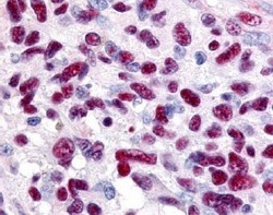
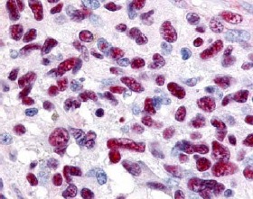
- Experimental details
- Immunohistochemistry: AP09260PU-N Human Gli-3 antibody was used at a 0.625 μg/ml to detect Gli-3 in a variety of tissues. Strong nuclear and smooth muscle staining was noted to be consistent with previously published reports. Specific staining was noted in tissue from adrenal, brain, glioblastoma, colon, heart, kidney, lung, liver, skeletal muscle, ovary, pancreas, placenta, skin, spleen, stomach, testes, thymus, thyroid, tonsil and uterus. This image shows Gli-3 staining of Human glioblastoma. Tissue was Formalin-Fixed and Paraffin Embedded.