Explore
 Validate
 Learn
Learn Western blot
Antibody data
- Antibody Data
- Antigen structure
- References [0]
- Comments [0]
- Validations
- Western blot [3]
- Immunohistochemistry [3]
Submit
Validation data
Reference
Comment
Report error
- Product number
- MA5-44726 - Provider product page

- Provider
- Invitrogen Antibodies
- Product name
- EMC4 Recombinant Rabbit Monoclonal Antibody (JE65-17)
- Antibody type
- Monoclonal
- Antigen
- Synthetic peptide
- Reactivity
- Human, Mouse, Rat
- Host
- Rabbit
- Isotype
- IgG
- Antibody clone number
- JE65-17
- Vial size
- 100 µL
- Concentration
- 1 mg/mL
- Storage
- Store at 4°C short term. For long term storage, store at -20°C, avoiding freeze/thaw cycles.
No comments: Submit comment
Supportive validation
- Submitted by
- Invitrogen Antibodies (provider)
- Main image

- Experimental details
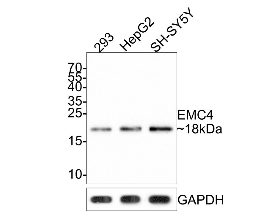
- Western blot analysis of EMC4 in different lysates (10 µg/Lane). Exposure time: 2 minutes; 15% SDS-PAGE gel. Proteins were transferred to a PVDF membrane and blocked with 5% NFDM/TBST for 1 hour at room temperature. Samples were incubated in EMC4 Monoclonal antibody (Product # MA5-44726) using a dilution of 1:500 in 5% NFDM/TBST at room temperature for 2 hours followed by Goat Anti-Rabbit IgG - HRP secondary antibody at a dilution of 1:300,000 for 1 hour at room temperature. Lane 1: 293 cell lysate; Lane 2: HepG2 cell lysate; Lane 3: SH-SY5Y cell lysate. Predicted band size: 20 kDa. Observed band size: 18 kDa.
- Submitted by
- Invitrogen Antibodies (provider)
- Main image

- Experimental details
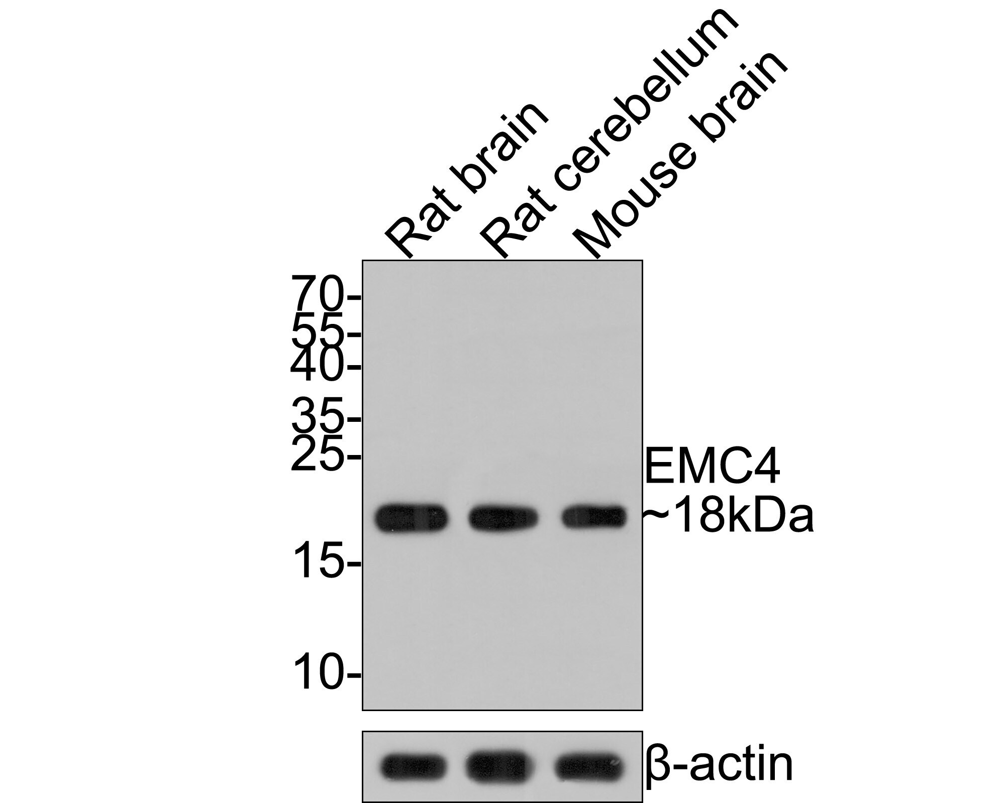
- Western blot analysis of EMC4 in different lysates (20 µg/Lane). Exposure time: 1 minute; 15% SDS-PAGE gel. Proteins were transferred to a PVDF membrane and blocked with 5% NFDM/TBST for 1 hour at room temperature. Samples were incubated in EMC4 Monoclonal antibody (Product # MA5-44726) using a dilution of 1:500 in 5% NFDM/TBST at room temperature for 2 hours followed by Goat Anti-Rabbit IgG - HRP secondary antibody at a dilution of 1:300,000 for 1 hour at room temperature. Lane 1: Rat brain tissue lysate; Lane 2: Rat cerebellum tissue lysate; Lane 3: Mouse brain tissue lysate. Predicted band size: 20 kDa. Observed band size: 18 kDa.
- Submitted by
- Invitrogen Antibodies (provider)
- Main image

- Experimental details
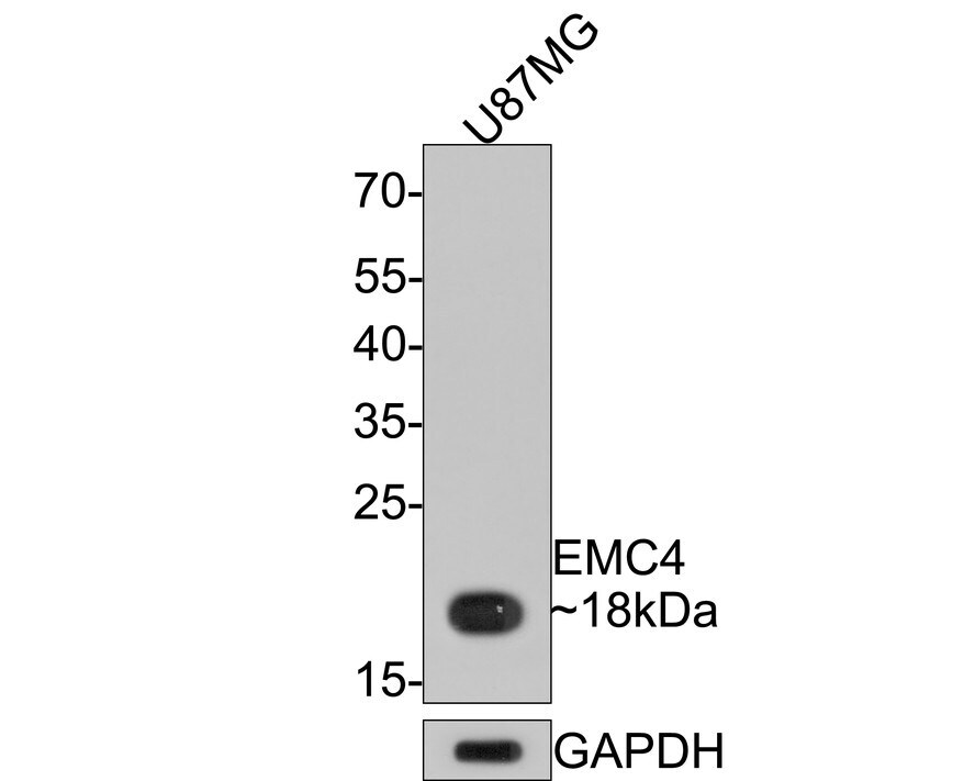
- Western blot analysis of EMC4 in U87MG cell lysates (10 µg/Lane). Exposure time: 30 seconds; 12% SDS-PAGE gel. Proteins were transferred to a PVDF membrane and blocked with 5% NFDM/TBST for 1 hour at room temperature. Samples were incubated in EMC4 Monoclonal antibody (Product # MA5-44726) using a dilution of 1:5,000 in 5% NFDM/TBST at room temperature for 2 hours followed by Goat Anti-Rabbit IgG - HRP secondary antibody at a dilution of 1:300,000 for 1 hour at room temperature. Predicted band size: 20 kDa. Observed band size: 18 kDa.
Supportive validation
- Submitted by
- Invitrogen Antibodies (provider)
- Main image
- Experimental details
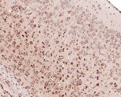
- Immunohistochemistry analysis of EMC4 in paraffin-embedded mouse brain tissue. The section was pre-treated using heat mediated antigen retrieval with Tris-EDTA buffer (pH 9.0) for 20 minutes. The tissues were blocked in 1% BSA for 20 minutes at room temperature, washed with ddH2O and PBS, and then probed with EMC4 Monoclonal antibody (Product # MA5-44726) using a dilution of 1:400 for 1 hour at room temperature. The detection was performed using an HRP conjugated compact polymer system. DAB was used as the chromogen. Tissues were counterstained with hematoxylin and mounted with DPX.

- Submitted by
- Invitrogen Antibodies (provider)
- Main image

- Experimental details
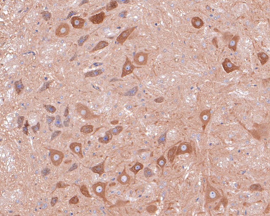
- Immunohistochemistry analysis of EMC4 in paraffin-embedded rat brain tissue. The section was pre-treated using heat mediated antigen retrieval with Tris-EDTA buffer (pH 9.0) for 20 minutes. The tissues were blocked in 1% BSA for 20 minutes at room temperature, washed with ddH2O and PBS, and then probed with EMC4 Monoclonal antibody (Product # MA5-44726) using a dilution of 1:400 for 1 hour at room temperature. The detection was performed using an HRP conjugated compact polymer system. DAB was used as the chromogen. Tissues were counterstained with hematoxylin and mounted with DPX.
- Submitted by
- Invitrogen Antibodies (provider)
- Main image

- Experimental details
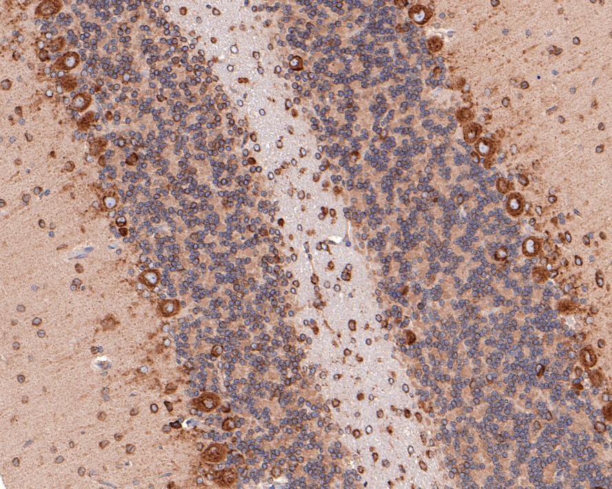
- Immunohistochemistry analysis of EMC4 in paraffin-embedded rat cerebellum tissue. The section was pre-treated using heat mediated antigen retrieval with Tris-EDTA buffer (pH 9.0) for 20 minutes. The tissues were blocked in 1% BSA for 20 minutes at room temperature, washed with ddH2O and PBS, and then probed with EMC4 Monoclonal antibody (Product # MA5-44726) using a dilution of 1:100 for 1 hour at room temperature. The detection was performed using an HRP conjugated compact polymer system. DAB was used as the chromogen. Tissues were counterstained with hematoxylin and mounted with DPX.