 Explore
 Validate
 Learn
Learn Western blot
Antibody data
- Antibody Data
- Antigen structure
- References [2]
- Comments [0]
- Validations
- Western blot [7]
- Immunocytochemistry [1]
- Immunohistochemistry [4]
- Flow cytometry [1]
- Other assay [3]
Submit
Validation data
Reference
Comment
Report error
- Product number
- PA5-19910 - Provider product page

- Provider
- Invitrogen Antibodies
- Product name
- CX3CR1 Polyclonal Antibody
- Antibody type
- Polyclonal
- Antigen
- Synthetic peptide
- Description
- In Western blot applications, this antibody detects a band at ~50kDa. A suggested positive control is human spleen tissue lysate. PA5-19910 can be used with blocking peptide PEP-0036.
- Reactivity
- Human
- Host
- Rabbit
- Isotype
- IgG
- Vial size
- 100 µg
- Concentration
- 1 mg/mL
- Storage
- Maintain refrigerated at 2-8°C for up to 3 months. For long term storage store at -20°C
Submitted references Validation of Induced Microglia-Like Cells (iMG Cells) for Future Studies of Brain Diseases.
Insights Into the Somatic Mutation Burden of Hepatoblastomas From Brazilian Patients.
Banerjee A, Lu Y, Do K, Mize T, Wu X, Chen X, Chen J
Frontiers in cellular neuroscience 2021;15:629279
Frontiers in cellular neuroscience 2021;15:629279
Insights Into the Somatic Mutation Burden of Hepatoblastomas From Brazilian Patients.
Aguiar TFM, Rivas MP, Costa S, Maschietto M, Rodrigues T, Sobral de Barros J, Barbosa AC, Valieris R, Fernandes GR, Bertola DR, Cypriano M, Caminada de Toledo SR, Major A, Tojal I, Apezzato MLP, Carraro DM, Rosenberg C, Lima da Costa CM, Cunha IW, Sarabia SF, Terrada DL, Krepischi ACV
Frontiers in oncology 2020;10:556
Frontiers in oncology 2020;10:556
No comments: Submit comment
Supportive validation
- Submitted by
- Invitrogen Antibodies (provider)
- Main image

- Experimental details
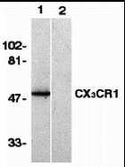
- Western blot analysis of human spleen lysate using a CX3CR1 polyclonal antibody (Product # PA5-19910) in the absence (lane 1) or presence of blocking peptide (lane 2).

- Submitted by
- Invitrogen Antibodies (provider)
- Main image

- Experimental details
- Western Blot Validation in Human Spleen Lysates. Loading: 15 µg of lysates per lane. Antibodies: CX3CR1 Polyclonal Antibody (Product # PA5-19910) (1 µg/mL) in the absence (lane 1) or presence of blocking peptide (lane 2), 1h incubation at RT in 0.05 NFDM/TBST. Secondary: Goat anti-rabbit IgG HRP conjugate at 1:10,000 dilution.
- Submitted by
- Invitrogen Antibodies (provider)
- Main image
- Experimental details
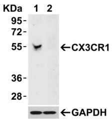
- Western Blot analysis of CX3CR1 in 293 cells. Lysates (15 µg) were loaded onto SDS-PAGE and blots were probed with CX3CR1 Polyclonal Antibody (Product # PA5-19910) diluted to 0.5 µg/mL. 1h incubation at RT in 0.05 NFDM/TBST. Secondary: Goat anti-rabbit IgG HRP conjugate at 1:10,000 dilution. Lane 1: 293 cells transfected with control siRNAs. Lane 2: 293 cells transfected with CX3CR1 siRNAs.

- Submitted by
- Invitrogen Antibodies (provider)
- Main image

- Experimental details
- Western Blot Validation in Human Cells. Loading: 15 µg of lysates per lane. Antibodies: CX3CR1 Polyclonal Antibody (Product # PA5-19910) (0.5 µg/mL), 1h incubation at RT in 0.05 NFDM/TBST. Secondary: Goat anti-rabbit IgG HRP conjugate at 1:10,000 dilution.

- Submitted by
- Invitrogen Antibodies (provider)
- Main image

- Experimental details
- Western Blot Validation in Human Spleen Lysates. Loading: 15 µg of lysates per lane. Antibodies: CX3CR1 Polyclonal Antibody (Product # PA5-19910) (1 µg/mL) in the absence (lane 1) or presence of blocking peptide (lane 2), 1h incubation at RT in 0.05 NFDM/TBST. Secondary: Goat anti-rabbit IgG HRP conjugate at 1:10,000 dilution.

- Submitted by
- Invitrogen Antibodies (provider)
- Main image

- Experimental details
- Western Blot Validation in Rat Spleen Tissue. Loading: 15 µg of lysates per lane. Antibodies: CX3CR1 Polyclonal Antibody (Product # PA5-19910) (A; 1 µg/mL, B; 2 µg/mL), 1h incubation at RT in 0.05 NFDM/TBST. Secondary: Goat anti-rabbit IgG HRP conjugate at 1:10,000 dilution.

- Submitted by
- Invitrogen Antibodies (provider)
- Main image
- Experimental details
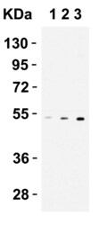
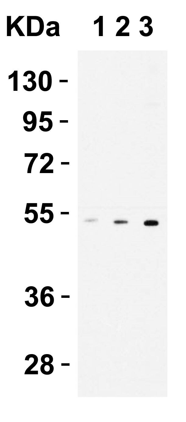
- Western Blot Validation in THP1 Cells. Loading: 15 µg of lysates per lane. Antibodies: CX3CR1 Polyclonal Antibody (Product # PA5-19910) (1 µg/mL), 1h incubation at RT in 0.05 NFDM/TBST. Secondary: Goat anti-rabbit IgG HRP conjugate at 1:10,000 dilution. Lane 1: 0.2 µg/mL Lane 1: 0.5 µg/mL Lane 1: 1 µg/mL
Supportive validation
- Submitted by
- Invitrogen Antibodies (provider)
- Main image

- Experimental details
- Immunofluorescent analysis of 4% paraformaldehyde-fixed K562 cells labeling CX3CR1 with CX3CR1 Polyclonal Antibody (Product # PA5-19910) at 10 µg/mL, followed by goat anti-rabbit IgG secondary antibody at 1:500 dilution (red) and DAPI staining (blue). Image showing membrane staining on K562 cells.

Supportive validation
- Submitted by
- Invitrogen Antibodies (provider)
- Main image

- Experimental details
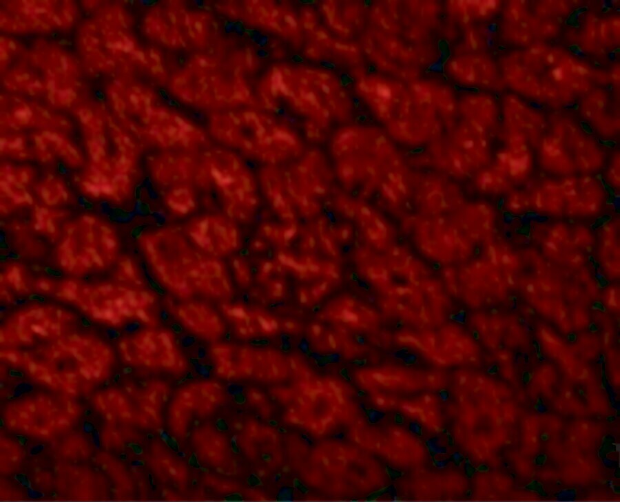
- Immunofluorescent analysis of 4% paraformaldehyde-fixed human heart tissue labeling CX3CR1 with CX3CR1 Polyclonal Antibody (Product # PA5-19910) at 10 µg/mL, followed by goat anti-rabbit IgG secondary antibody at 1:500 dilution (red).
- Submitted by
- Invitrogen Antibodies (provider)
- Main image

- Experimental details
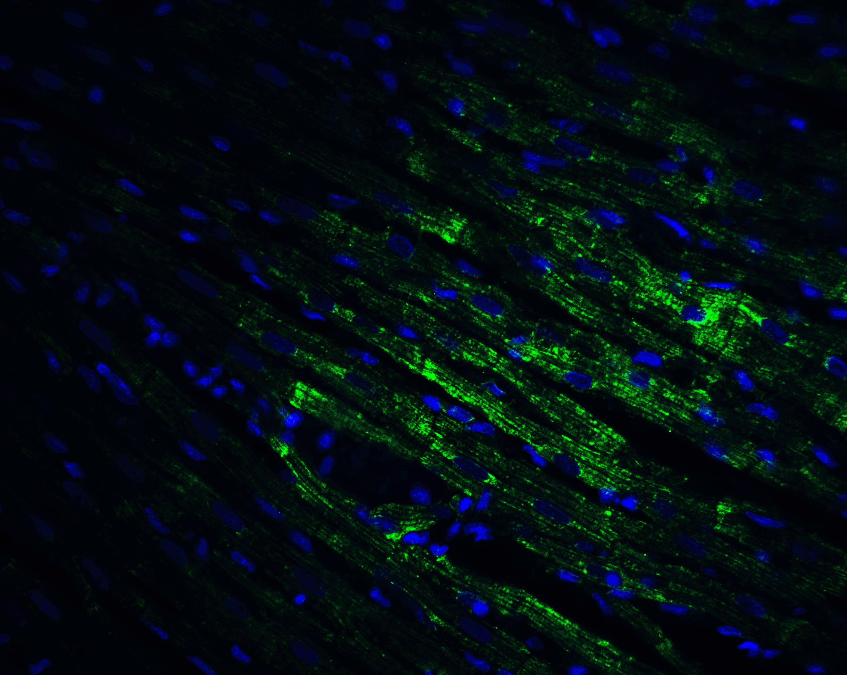
- Immunofluorescent analysis of 4% paraformaldehyde-fixed mouse heart tissue labeling CX3CR1 with CX3CR1 Polyclonal Antibody (Product # PA5-19910) at 20 µg/mL, followed by goat anti-rabbit IgG secondary antibody at 1:500 dilution (green) and DAPI staining (blue).

- Submitted by
- Invitrogen Antibodies (provider)
- Main image

- Experimental details
- Immunofluorescent analysis of 4% paraformaldehyde-fixed rat heart tissue labeling CX3CR1 with CX3CR1 Polyclonal Antibody (Product # PA5-19910) at 20 µg/mL, followed by goat anti-rabbit IgG secondary antibody at 1:500 dilution (green) and DAPI staining (blue).
- Submitted by
- Invitrogen Antibodies (provider)
- Main image

- Experimental details
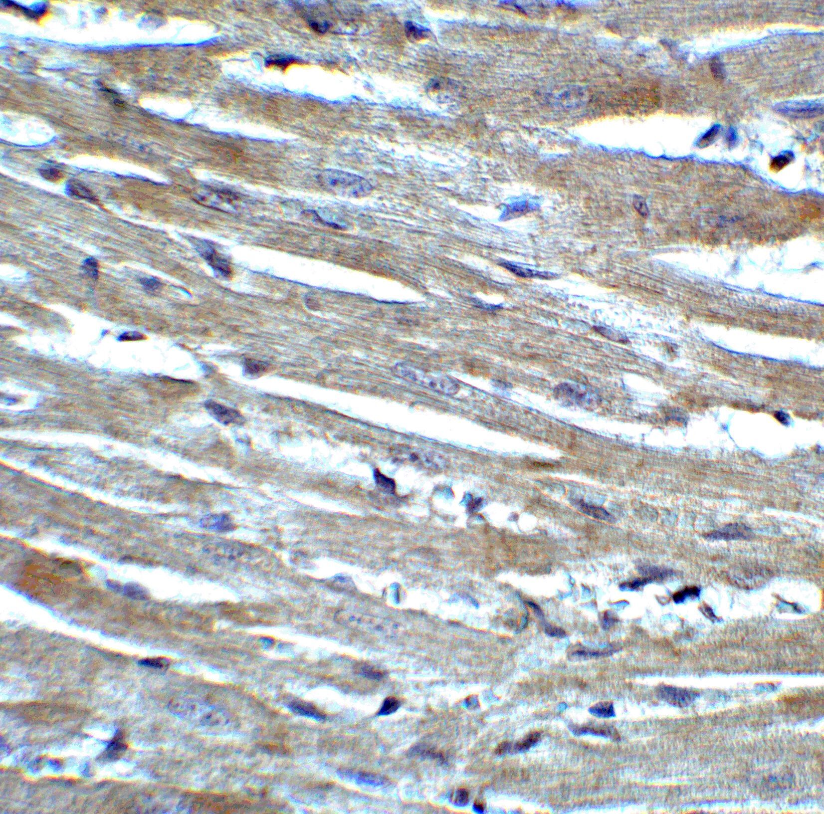
- Immunohistochemical analysis of paraffin-embedded rat heart tissue using CX3CR1 Polyclonal Antibody (Product # PA5-19910) at 2 µg/mL. Tissue was fixed with formaldehyde and blocked with 0.1 serum for 1 h at RT; antigen retrieval was by heat mediation with a citrate buffer (pH6). Samples were incubated with primary antibody overnight at 4˚C. A goat anti-rabbit IgG H&L (HRP) at 1/250 was used as secondary. Counter stained with Hematoxylin.
Supportive validation
- Submitted by
- Invitrogen Antibodies (provider)
- Main image

- Experimental details
- Flow Cytometry Validation of CX3CR1 in THP-1 Cells Overlay histogram showing THP-1 cells stained with CX3CR1 Polyclonal Antibody (Product # PA5-19910) (red line, 1 µg/1 x 10^6 cells). 1 h incubation at 4°C in 2% FBS/PBS. Followed by secondary antibody 488 goat anti-rabbit IgG (H+L) at 1/500 dilution for 1 h 4°C. Isotype control antibody (Green line) was rabbit IgG1 (1 µg/1 x 10^6 cells) used under the same conditions. Acquisition of >10,000 events was performed.

Supportive validation
- Submitted by
- Invitrogen Antibodies (provider)
- Main image
- Experimental details
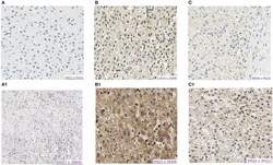
- Figure 3 Protein expression of CX3CL1 and CX3CR1 evaluated in hepatoblastoma samples by immunohistochemistry assay. (A-C) Show CX3CR1 data, and (A1-C1) , CX3CL1 from the same tumor samples. (A) HB17, example of negative labeling for CX3CR1 (A) and CX3CL1 (A1) . (B) HB32T, positive for nuclear and cytoplasmic CX3CR1 (B) and CX3CL1 (B1) . (C) HB33T, positive for cytoplasmic CX3CR1 labeling (C) and positive for nuclear and cytoplasmic CX3CL1 (C1) .

- Submitted by
- Invitrogen Antibodies (provider)
- Main image

- Experimental details
- Figure 4 Protein expression of CX3CL1 and CX3CR1 evaluated in hepatoblastomas and hepatoblastoma lung metastasis by immunohistochemistry assay. (A-D) Show CX3CL1 data, and (A1-D1) , CX3CR1. (A) TCH361, CX3CL1 strong positivity of infiltrated lymphocytes (indicated by arrow 1) in necrotic regions of the tumor, and (A1) , CX3CR1 strong positivity of infiltrated lymphocytes (indicated by arrow 2) in necrotic regions of the tumor; (B,B1) TCH327, positivity in tumor cells (indicated by arrows 3 and 5) and infiltrated lymphocytes negative (indicated by arrows 4 and 6) for both proteins. (C) TCH321, positivity in the osteoblast component and strong positivity in the fetal type (indicated by arrow 7); infiltrated lymphocytes are negative (indicated by arrow 8); (C1) positivity in tumor cells and lymphocytes negative; (D,D1) TCH360, lung metastasis showing positivity in tumor cells (indicated by arrows 9 and 11), and no expression in infiltrated lymphocytes (indicated by arrows 10 and 12), for both proteins.

- Submitted by
- Invitrogen Antibodies (provider)
- Main image
- Experimental details
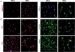
- Figure 2 iMG cells expressed microglia-specific markers (TMEM119 and P2RY12) and other myeloid-specific markers. The expression of surface markers on the iMG cells and iMacs were performed by immunocytochemistry. Peripheral monocytes were incubated with GM-CSF (iMacs) for 5-7 days or cocktail of GM-CSF and IL-34 (iMG cells) for 10-14 days. Representative images of iMacs and iMG cells were shown. (A) iMG cells expressed unique microglial markers TMEM119 (b) and P2RY12 (d), which were absent from iMacs (a,c). (B) Myeloid lineage markers were expressed on both iMacs and iMG cells, such as Iba1 (a,b), PU.1 (c,d), CX3CR1 (e,f), and TREM2 (g,h). Images were captured at 40x with a scale representing 20 mum, where P2RY12, CX3CR1, and TREM2 were stained green, TMEM119, Iba1, and PU.1 were stained red. Cellular nuclei were stained blue with DAPI.
