 Explore
 Validate
 Learn
Learn Western blot
Antibody data
- Antibody Data
- Antigen structure
- References [2]
- Comments [0]
- Validations
- Western blot [2]
- Immunocytochemistry [1]
- Immunohistochemistry [3]
Submit
Validation data
Reference
Comment
Report error
- Product number
- GTX111934 - Provider product page

- Provider
- GeneTex
- Proper citation
- GeneTex Cat#GTX111934, RRID:AB_1951755
- Product name
- RPL17 antibody [N1C3-2]
- Antibody type
- Polyclonal
- Reactivity
- Human, Rat
- Host
- Rabbit
Submitted references Human PDCD2L Is an Export Substrate of CRM1 That Associates with 40S Ribosomal Subunit Precursors.
Splicing-factor oncoprotein SRSF1 stabilizes p53 via RPL5 and induces cellular senescence.
Landry-Voyer AM, Bilodeau S, Bergeron D, Dionne KL, Port SA, Rouleau C, Boisvert FM, Kehlenbach RH, Bachand F
Molecular and cellular biology 2016 Dec 15;36(24):3019-3032
Molecular and cellular biology 2016 Dec 15;36(24):3019-3032
Splicing-factor oncoprotein SRSF1 stabilizes p53 via RPL5 and induces cellular senescence.
Fregoso OI, Das S, Akerman M, Krainer AR
Molecular cell 2013 Apr 11;50(1):56-66
Molecular cell 2013 Apr 11;50(1):56-66
No comments: Submit comment
Supportive validation
- Submitted by
- GeneTex (provider)
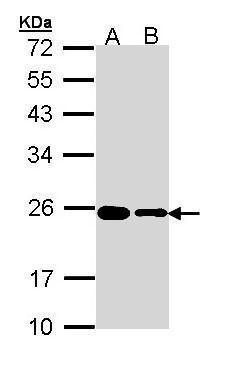
- Main image

- Experimental details
- Sample (30 ?g of whole cell lysate) A: HeLa B: HepG2 (GTX27900) 12% SDS PAGE GTX111934 diluted at 1:1000 The HRP-conjugated anti-rabbit IgG antibody (GTX213110-01) was used to detect the primary antibody.
- Submitted by
- GeneTex (provider)
- Main image
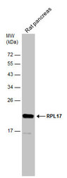
- Experimental details
- Rat tissue extract (50 ?g) was separated by 12% SDS-PAGE, and the membrane was blotted with RPL17 antibody [N1C3-2] (GTX111934) diluted at 1:1000. The HRP-conjugated anti-rabbit IgG antibody (GTX213110-01) was used to detect the primary antibody.

Supportive validation
- Submitted by
- GeneTex (provider)
- Main image
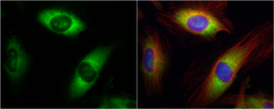
- Experimental details
- RPL17 antibody [N1C3-2] detects RPL17 protein at cytoplasm by immunofluorescent analysis.Sample: HeLa cells were fixed in 4% paraformaldehyde at RT for 15 min.Green: RPL17 protein stained by RPL17 antibody [N1C3-2] (GTX111934) diluted at 1:500.Red: alpha Tubulin, a cytoskeleton marker, stained by alpha Tubulin antibody [B-5-1-2] (GTX11304) diluted at 1:10000.Blue: Hoechst 33342 staining.

Supportive validation
- Submitted by
- GeneTex (provider)
- Main image
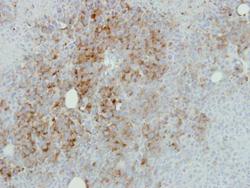
- Experimental details
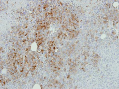
- Immunohistochemical analysis of paraffin-embedded HBL435 xenograft, using RPL17(GTX111934) antibody at 1:500 dilution.
- Submitted by
- GeneTex (provider)
- Main image
- Experimental details
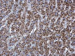
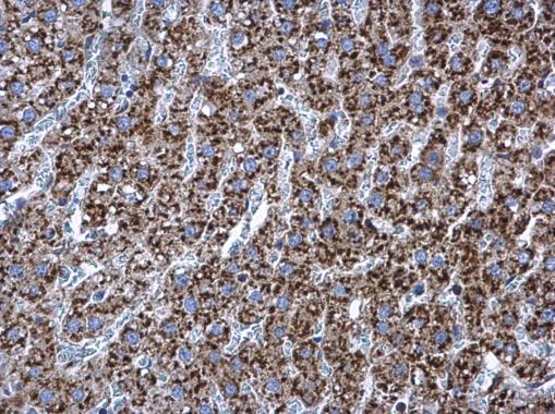
- RPL17 antibody [N1C3-2] detects RPL17 protein at cytoplasm in rat liver by immunohistochemical analysis. Sample: Paraffin-embedded rat liver. RPL17 antibody [N1C3-2] (GTX111934) diluted at 1:500.
- Submitted by
- GeneTex (provider)
- Main image
- Experimental details
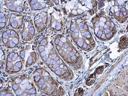
- RPL17 antibody [N1C3-2] detects RPL17 protein at cytoplasm in rat colon by immunohistochemical analysis. Sample: Paraffin-embedded rat colon. RPL17 antibody [N1C3-2] (GTX111934) diluted at 1:500.
