Explore
 Validate
 Learn
Learn Immunocytochemistry
Antibody data
- Antibody Data
- Antigen structure
- References [1]
- Comments [0]
- Validations
- Immunocytochemistry [1]
- Immunohistochemistry [5]
- Other assay [1]
Submit
Validation data
Reference
Comment
Report error
- Product number
- MA5-31360 - Provider product page

- Provider
- Invitrogen Antibodies
- Product name
- ATF3 Monoclonal Antibody (CL1685)
- Antibody type
- Monoclonal
- Antigen
- Recombinant full-length protein
- Description
- Immunogen sequence: MMLQHPGQVS ASEVSASAIV PCLSPPGSLV FEDFANLTPF VKEELRFAIQ NKHLCHRMSS ALESVTVSDR PLGVSITKAE VAPEEDERKK RRRERNKIAA AKCRNKKKEK TEC
- Reactivity
- Human
- Host
- Mouse
- Isotype
- IgG
- Antibody clone number
- CL1685
- Vial size
- 100 µL
- Concentration
- 1 mg/mL
- Storage
- Store at 4°C short term. For long term storage, store at -20°C, avoiding freeze/thaw cycles.
Submitted references Profound gene expression changes in the epithelial monolayer of active ulcerative colitis and Crohn's disease.
Sæterstad S, Østvik AE, Røyset ES, Bakke I, Sandvik AK, Granlund AVB
PloS one 2022;17(3):e0265189
PloS one 2022;17(3):e0265189
No comments: Submit comment
Supportive validation
- Submitted by
- Invitrogen Antibodies (provider)
- Main image

- Experimental details
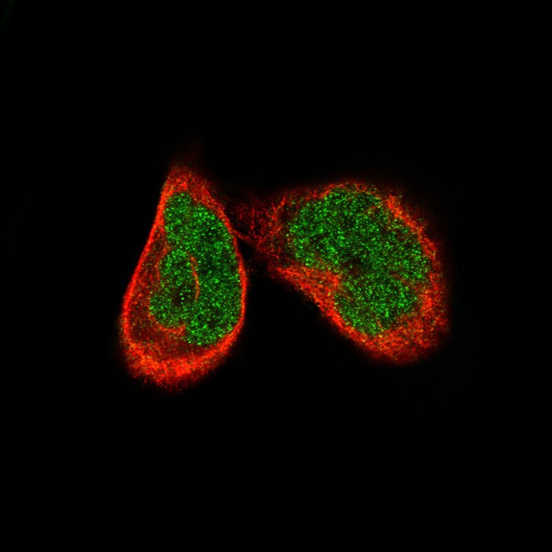
- Immunocytochemistry-Immunofluorescence analysis of ATF3 in RH-30 cells using ATF3 Monoclonal Antibody (CL1685) (Product # MA5-31360), showing specific staining in the nucleoplasm in green. Microtubule- and nuclear probes are visualized in red and blue, respectively (where available).
Supportive validation
- Submitted by
- Invitrogen Antibodies (provider)
- Main image
- Experimental details
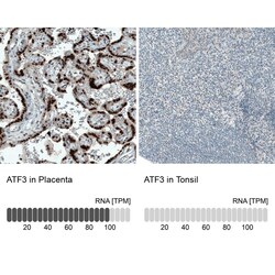
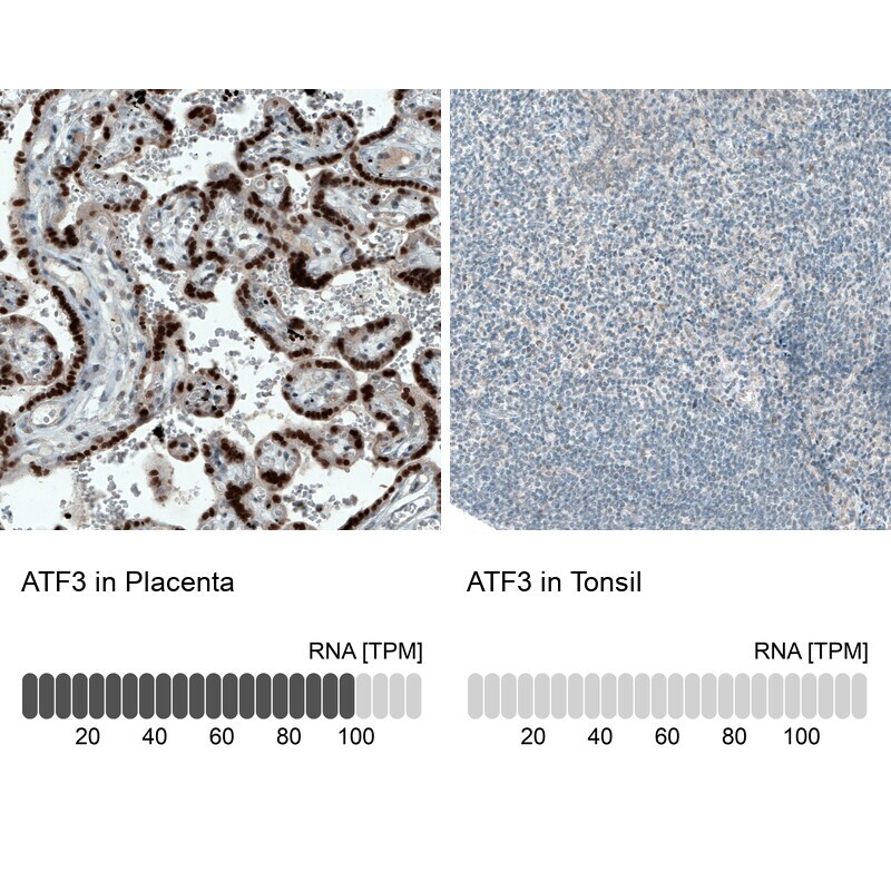
- Immunohistochemical analysis of ATF3 in human placenta and tonsil tissues using a ATF3 monoclonal antibody (Product # MA5-31360). Corresponding RNA-seq data are presented for the same tissues.
- Submitted by
- Invitrogen Antibodies (provider)
- Main image
- Experimental details
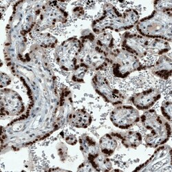
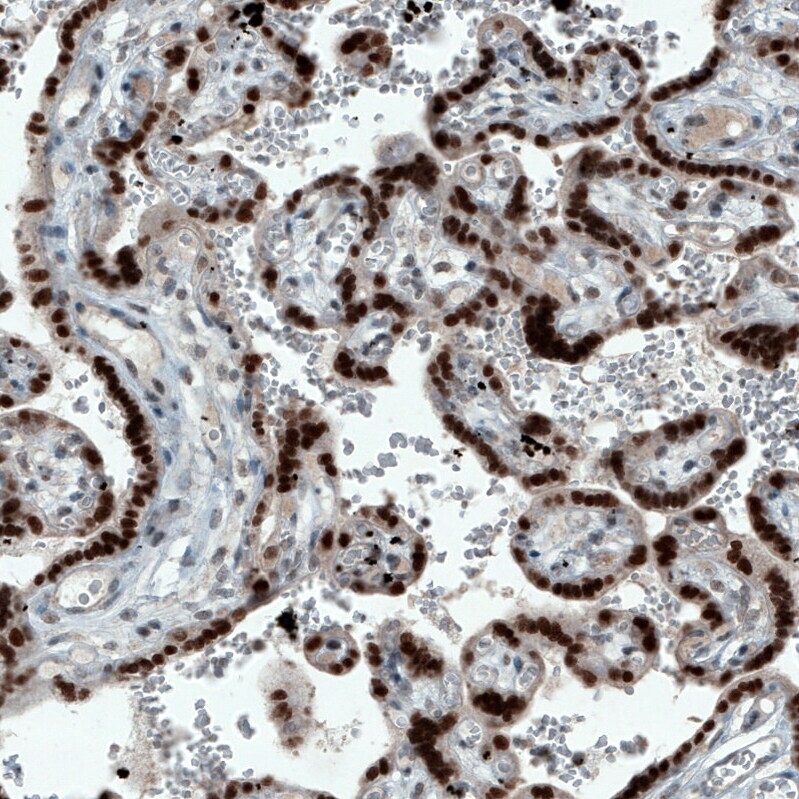
- Immunohistochemical analysis of ATF3 in human placenta using a ATF3 monoclonal antibody (Product # MA5-31360). The analysis shows strong nuclear positivity in trophoblastic cells.
- Submitted by
- Invitrogen Antibodies (provider)
- Main image
- Experimental details
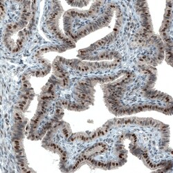
- Immunohistochemical analysis of ATF3 in human fallopian tube using a ATF3 monoclonal antibody (Product # MA5-31360). The analysis shows moderate to strong nuclear positivity in glandular cells.

- Submitted by
- Invitrogen Antibodies (provider)
- Main image

- Experimental details
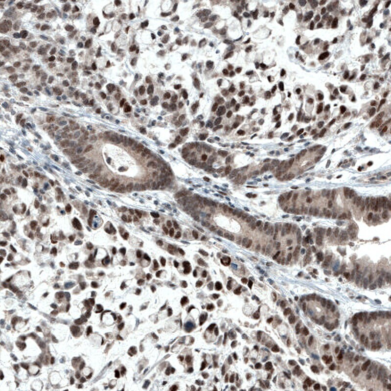
- Immunohistochemical analysis of ATF3 in human stomach cancer using a ATF3 monoclonal antibody (Product # MA5-31360). The analysis shows moderate to strong nuclear positivity in tumor cells.
- Submitted by
- Invitrogen Antibodies (provider)
- Main image
- Experimental details
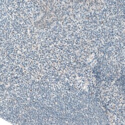
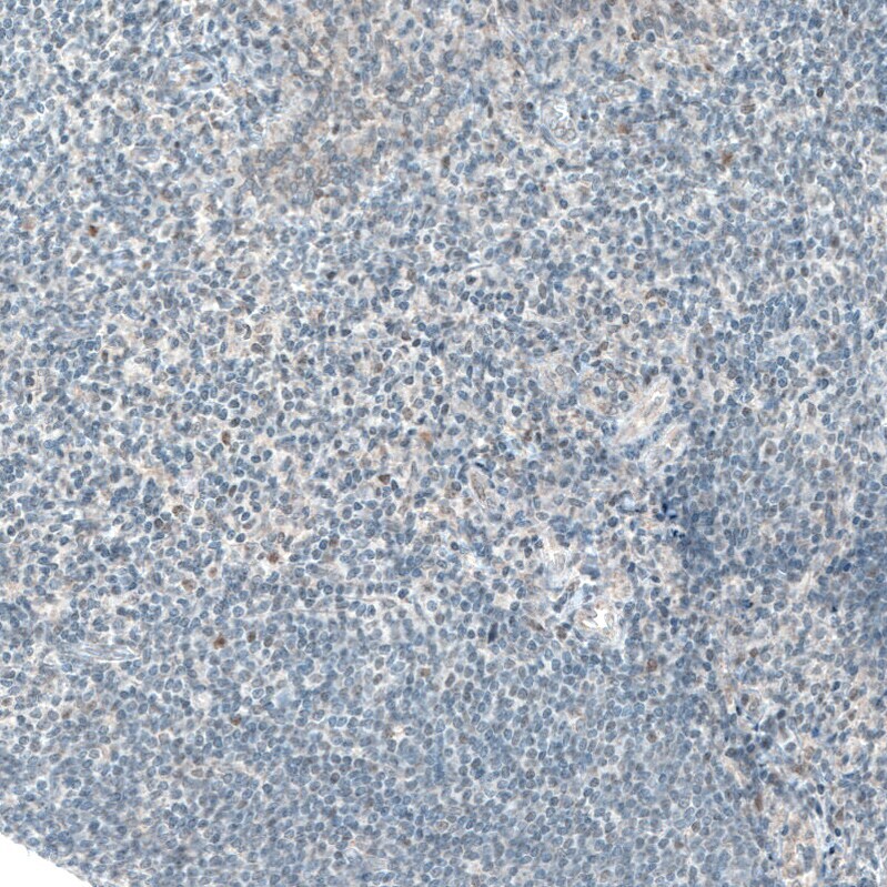
- Immunohistochemical analysis of ATF3 in human tonsil using a ATF3 monoclonal antibody (Product # MA5-31360). The analysis shows no positivity in lymphoid cells as expected.
Supportive validation
- Submitted by
- Invitrogen Antibodies (provider)
- Main image
- Experimental details
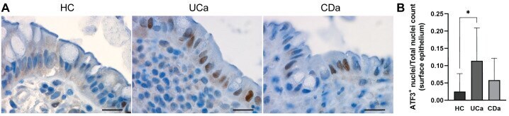
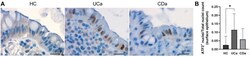
- 10.1371/journal.pone.0265189.g006 Fig 6 Immunohistochemical ATF3-quantitation confirmed increased nuclear ATF3 positivity in UCa surface epithelium compared to HC. ( A ) Representative ATF3 staining from HC (left), UCa (middle) and CDa (right). Scale bars (20mum) indicated. ( B ) A series of sections from HC ( n = 12), UCa ( n = 11) and CDa ( n = 8) were quantified using QuPath. For each section, surface epithelial ATF3-positivity is presented as the ratio of ATF3 + nuclei and total nuclei count. One-way ANOVA followed by Dunnett's multiple comparisons test showed that nuclear ATF3 positivity was significantly higher in UCa than HC (adj. p = 0.013), but not in CDa vs. HC (adj. p = 0.521).