Explore
 Validate
 Learn
Learn Western blot
Antibody data
- Antibody Data
- Antigen structure
- References [1]
- Comments [0]
- Validations
- Western blot [3]
- Immunocytochemistry [2]
- Immunohistochemistry [2]
- Other assay [2]
Submit
Validation data
Reference
Comment
Report error
- Product number
- PA5-28369 - Provider product page

- Provider
- Invitrogen Antibodies
- Product name
- HSPA1B Polyclonal Antibody
- Antibody type
- Polyclonal
- Antigen
- Recombinant protein fragment
- Description
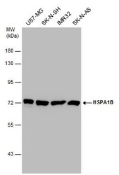
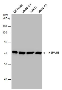
- Recommended positive controls: U87-MG, SK-N-SH, IMR32, SK-N-AS.
- Concentration
- 0.55 mg/mL
Submitted references Deletion of the Capn1 Gene Results in Alterations in Signaling Pathways Related to Alzheimer's Disease, Protein Quality Control and Synaptic Plasticity in Mouse Brain.
Su W, Zhou Q, Wang Y, Chishti A, Li QQ, Dayal S, Shiehzadegan S, Cheng A, Moore C, Bi X, Baudry M
Frontiers in genetics 2020;11:334
Frontiers in genetics 2020;11:334
No comments: Submit comment
Supportive validation
- Submitted by
- Invitrogen Antibodies (provider)
- Main image

- Experimental details
- Western blot analysis of HSP70/Heat Shock Protein 70 using 30 µg of A) 293T (B) A431 and C) H1299 lysate. Samples were loaded onto a 12% SDS-PAGE gel and probed with a HSP70/Heat Shock Protein 70 polyclonal antibody (Product # PA5-28369) at a dilution of 1:500.
- Submitted by
- Invitrogen Antibodies (provider)
- Main image

- Experimental details
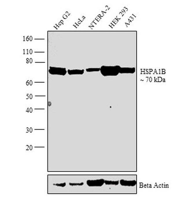
- Western blot analysis was performed on whole cell extracts (30 µg lysate) of Hep G2 (Lane 1), HeLa (Lane 2), NTERA-2 (Lane 3), HEK 293 (Lane 4) and A431 (Lane 5). The blot was probed with Anti- HSPA1B Polyclonal Antibody (Product # PA5-28369, 1 µg/ml) and detected by chemiluminescence using Goat anti-Rabbit IgG (H+L) Superclonal™ Secondary Antibody, HRP conjugate (Product # A27036, 0.25 µg/ml, 1:4000 dilution). A 70 kDa band corresponding to HSPA1B was observed across all the cell lines tested.
- Submitted by
- Invitrogen Antibodies (provider)
- Main image
- Experimental details
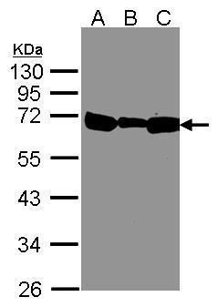
- Western Blot analysis of HSPA1B was performed by separating 30 µg of various whole cell extracts by 7.5% SDS-PAGE. Proteins were transferred to a membrane and probed with a HSPA1B Polyclonal Antibody (Product # PA5-28369) at a dilution of 1:2000 and a HRP-conjugated anti-rabbit IgG secondary antibody.
Supportive validation
- Submitted by
- Invitrogen Antibodies (provider)
- Main image

- Experimental details
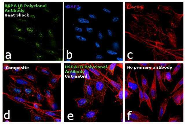
- Immunofluorescence analysis of HSPA1B was performed using 70% confluent log phase PC-3 cells treated with Heat Shock (43 degree celsius for 1 hr). The cells were fixed with 4% paraformaldehyde for 10 minutes, permeabilized with 0.1% Triton™ X-100 for 15 minutes, and blocked with 1% BSA for 1 hour at room temperature. The cells were labeled with HSPA1B Polyclonal Antibody (Product # PA5-28369) at 5 µg/mL concentration in 0.1% BSA, incubated at 4 degree celsius overnight and then labeled with Goat anti-Rabbit IgG (H+L) Superclonal™ Secondary Antibody, Alexa Fluor® 488 conjugate (Product # A27034) at a dilution of 1:2000 for 45 minutes at room temperature (Panel a: green). Nuclei (Panel b: blue) were stained with ProLong™ Diamond Antifade Mountant with DAPI (Product # P36962). F-actin (Panel c: red) was stained with Rhodamine Phalloidin (Product # R415, 1:300). Panel d represents the merged image showing HSPA1B predominantly in the nucleolus and faint in the cytoplasm. Panel e represents the untreated cells showing faint cytoplasmic and nucleus localization. Panel f represents control cells with no primary antibody to assess background. The images were captured at 60X magnification.
- Submitted by
- Invitrogen Antibodies (provider)
- Main image
- Experimental details
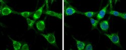
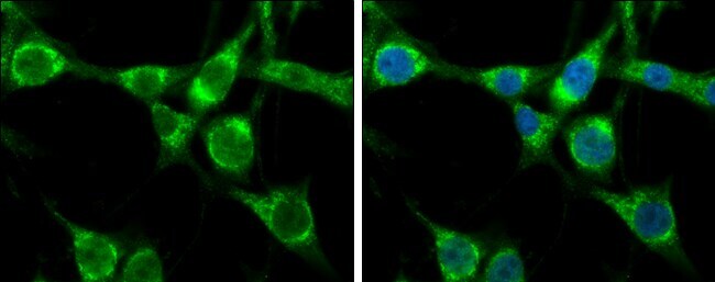
- Immunocytochemistry-Immunofluorescence analysis of HSPA1B was performed in U-87 MG cells fixed in 4% paraformaldehyde at RT for 15 min. Green: HSPA1B Polyclonal Antibody (Product # PA5-28369) diluted at 1:400. Blue: Hoechst 33342 staining.
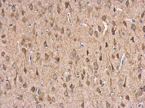
Supportive validation
- Submitted by
- Invitrogen Antibodies (provider)
- Main image
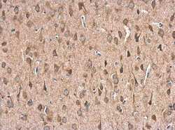
- Experimental details
- Immunohistochemistry (Paraffin) analysis of HSPA1B was performed in paraffin-embedded rat brain tissue using HSPA1B Polyclonal Antibody (Product # PA5-28369) at a dilution of 1:500.
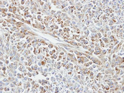
- Submitted by
- Invitrogen Antibodies (provider)
- Main image
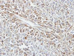
- Experimental details
- Immunohistochemical analysis of paraffin-embedded human glioma, using Hsp70 (Product # PA5-28369) antibody at 1:100 dilution. Antigen Retrieval: Citrate buffer, pH 6.0, 15 min.
Supportive validation
- Submitted by
- Invitrogen Antibodies (provider)
- Main image
- Experimental details
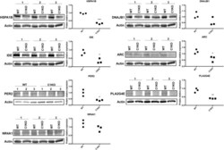
- FIGURE 4 RNA-seq results were validated by Western blot analysis in WT and calpain-1 KO mice. Western blot images and quantifications showing the expression levels of HSPA1B, DNAJB1, IDE, ARC, PER2, PLA2G4E, and NR4A1 proteins in three independent replicates, respectively. Dot plots were used for the quantification of the expression levels of 7 proteins compared to actin control in WT and calpain-1 KO mice. Unpaired t-test in Prism 7 was used to calculate p values, * p < 0.05, ** p < 0.01, *** p < 0.001.

- Submitted by
- Invitrogen Antibodies (provider)
- Main image
- Experimental details
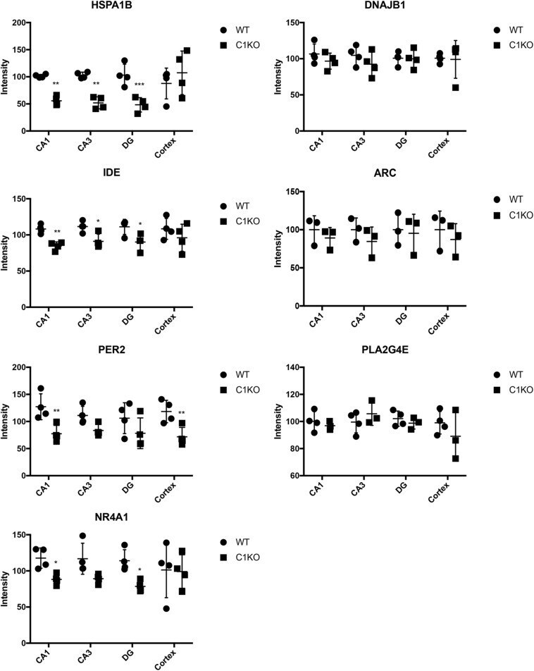
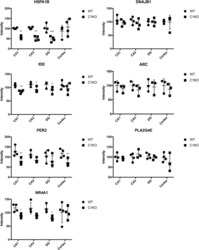
- FIGURE 5 Quantification of immunostaining for 7 proteins in the brain of WT and calpain-1 KO mice. Immunostaining for WT and calpain-1 KO mice was performed in brain slices. The mean fluorescence intensity was quantified in CA1, CA3, DG and cortex. Scatter dot plots were used to represent the expression of 7 proteins in four regions, with the lines in the dot plots representing standard deviations. Unpaired t-test in Prism 7 was used to calculate p values, * p < 0.05, ** p < 0.01, *** p < 0.001.