Antibody data
- Antibody Data
- Antigen structure
- References [4]
- Comments [0]
- Validations
- Western blot [5]
- Immunocytochemistry [2]
- Immunohistochemistry [2]
Submit
Validation data
Reference
Comment
Report error
- Product number
- GTX110587 - Provider product page

- Provider
- GeneTex
- Proper citation
- GeneTex Cat#GTX110587, RRID:AB_1952035
- Product name
- STAT3 antibody [C2C3], C-term
- Antibody type
- Polyclonal
- Reactivity
- Human, Mouse
- Host
- Rabbit
Submitted references Galectin-1 Reduces Neuroinflammation via Modulation of Nitric Oxide-Arginase Signaling in HIV-1 Transfected Microglia: a Gold Nanoparticle-Galectin-1 "Nanoplex" a Possible Neurotherapeutic?
Adipose-Derived Stem Cells Enhance Cancer Stem Cell Property and Tumor Formation Capacity in Lewis Lung Carcinoma Cells Through an Interleukin-6 Paracrine Circuit.
The significance of microRNA-184 on JAK2/STAT3 signaling pathway in the formation mechanism of glioblastoma.
Inhibitory effect of litchi (Litchi chinensis Sonn.) flower on lipopolysaccharide-induced expression of proinflammatory mediators in RAW264.7 cells through NF-κB, ERK, and JAK2/STAT3 inactivation.
Aalinkeel R, Mangum CS, Abou-Jaoude E, Reynolds JL, Liu M, Sundquist K, Parikh NU, Chaves LD, Mammen MJ, Schwartz SA, Mahajan SD
Journal of neuroimmune pharmacology : the official journal of the Society on NeuroImmune Pharmacology 2017 Mar;12(1):133-151
Journal of neuroimmune pharmacology : the official journal of the Society on NeuroImmune Pharmacology 2017 Mar;12(1):133-151
Adipose-Derived Stem Cells Enhance Cancer Stem Cell Property and Tumor Formation Capacity in Lewis Lung Carcinoma Cells Through an Interleukin-6 Paracrine Circuit.
Lu JH, Wei HJ, Peng BY, Chou HH, Chen WH, Liu HY, Deng WP
Stem cells and development 2016 Dec 1;25(23):1833-1842
Stem cells and development 2016 Dec 1;25(23):1833-1842
The significance of microRNA-184 on JAK2/STAT3 signaling pathway in the formation mechanism of glioblastoma.
Zhang X, Ding H, Han Y, Sun D, Wang H, Zhai XU
Oncology letters 2015 Dec;10(6):3510-3514
Oncology letters 2015 Dec;10(6):3510-3514
Inhibitory effect of litchi (Litchi chinensis Sonn.) flower on lipopolysaccharide-induced expression of proinflammatory mediators in RAW264.7 cells through NF-κB, ERK, and JAK2/STAT3 inactivation.
Yang DJ, Chang YY, Lin HW, Chen YC, Hsu SH, Lin JT
Journal of agricultural and food chemistry 2014 Apr 16;62(15):3458-65
Journal of agricultural and food chemistry 2014 Apr 16;62(15):3458-65
No comments: Submit comment
Enhanced validation
Supportive validation
- Submitted by
- GeneTex (provider)
- Enhanced method
- Genetic validation
- Main image

- Experimental details
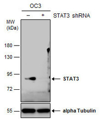
- Non-transfected (¡V) and transfected (+) OC3 whole cell extracts (30 ?g) were separated by 7.5% SDS-PAGE, and the membrane was blotted with STAT3 antibody [C2C3], C-term (GTX110587) diluted at 1:500. The HRP-conjugated anti-rabbit IgG antibody (GTX213110-01) was used to detect the primary antibody.
Supportive validation
- Submitted by
- GeneTex (provider)
- Main image

- Experimental details
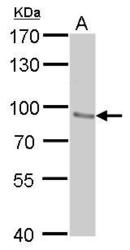
- STAT3 antibody detects STAT3 protein by western blot analysis.A. 30 ?g NIH-3T3 whole cell lysate/extract7.5% SDS-PAGESTAT3 antibody (GTX110587) dilution: 1:500 The HRP-conjugated anti-rabbit IgG antibody (GTX213110-01) was used to detect the primary antibody.
- Submitted by
- GeneTex (provider)
- Main image

- Experimental details
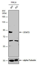
- Wild-type (WT) and STAT3 knockout (KO) HeLa cell extracts (30 ?g) were separated by 7.5% SDS-PAGE, and the membrane was blotted with STAT3 antibody [C2C3], C-term (GTX110587) diluted at 1:500. The HRP-conjugated anti-rabbit IgG antibody (GTX213110-01) was used to detect the primary antibody, and the signal was developed with Trident ECL plus-Enhanced.
- Submitted by
- GeneTex (provider)
- Main image

- Experimental details
- Non-transfected (¡V) and transfected (+) OC3 whole cell extracts (30 ?g) were separated by 7.5% SDS-PAGE, and the membrane was blotted with STAT3 antibody [C2C3], C-term (GTX110587) diluted at 1:500. The HRP-conjugated anti-rabbit IgG antibody (GTX213110-01) was used to detect the primary antibody.
- Submitted by
- GeneTex (provider)
- Main image

- Experimental details
- Wild-type (WT) and STAT3 knockout (KO) HeLa cell extracts (30 ?g) were separated by 7.5% SDS-PAGE, and the membrane was blotted with STAT3 antibody [C2C3], C-term (GTX110587) diluted at 1:500. The HRP-conjugated anti-rabbit IgG antibody (GTX213110-01) was used to detect the primary antibody, and the signal was developed with Trident ECL plus-Enhanced.
Supportive validation
- Submitted by
- GeneTex (provider)
- Main image

- Experimental details
- Immunofluorescence analysis of paraformaldehyde-fixed Human ESC, using STAT3(GTX110587) antibody at 1:200 dilution.
- Submitted by
- GeneTex (provider)
- Main image

- Experimental details
- Immunofluorescence analysis of paraformaldehyde-fixed HeLa, using STAT3(GTX110587) antibody at 1:200 dilution.
Supportive validation
- Submitted by
- GeneTex (provider)
- Main image

- Experimental details
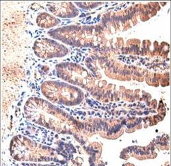
- Immunohistochemical analysis of paraffin-embedded mouse small intestine, using STAT3(GTX110587) antibody at 1:200 dilution.(Image courtesy of Koji Taniguchi, Ph.D (Laboratory of Dr. Michael Karin, UCSD.)
- Submitted by
- GeneTex (provider)
- Main image

- Experimental details
- Immunohistochemical analysis of paraffin-embedded NCIN87 xenograft, using STAT3(GTX110587) antibody at 1:100 dilution.
 Explore
 Validate
 Learn
Learn Western blot
 Immunocytochemistry
 Flow cytometry