 Explore
 Validate
 Learn
Learn Western blot
Antibody data
- Antibody Data
- Antigen structure
- References [2]
- Comments [0]
- Validations
- Western blot [2]
- Immunocytochemistry [3]
- Immunohistochemistry [6]
- Other assay [1]
Submit
Validation data
Reference
Comment
Report error
- Product number
- PA5-54849 - Provider product page

- Provider
- Invitrogen Antibodies
- Product name
- NECAB1 Polyclonal Antibody
- Antibody type
- Polyclonal
- Antigen
- Recombinant full-length protein
- Description
- Immunogen sequence: LLKETLNQLQ SLQNSLECAM ETTEEQTRQE RQGPAKPEVL SIQWPGKRSS RRVQRHNSFS PNSP
- Concentration
- 0.18 mg/mL
Submitted references Severe intraventricular hemorrhage causes long-lasting structural damage in a preterm rabbit pup model.
Behavioral defects associated with amygdala and cortical dysfunction in mice with seeded α-synuclein inclusions.
Romantsik O, Ross-Munro E, Grönlund S, Holmqvist B, Brinte A, Gerdtsson E, Vallius S, Bruschettini M, Wang X, Fleiss B, Ley D
Pediatric research 2022 Aug;92(2):403-414
Pediatric research 2022 Aug;92(2):403-414
Behavioral defects associated with amygdala and cortical dysfunction in mice with seeded α-synuclein inclusions.
Stoyka LE, Arrant AE, Thrasher DR, Russell DL, Freire J, Mahoney CL, Narayanan A, Dib AG, Standaert DG, Volpicelli-Daley LA
Neurobiology of disease 2020 Feb;134:104708
Neurobiology of disease 2020 Feb;134:104708
No comments: Submit comment
Supportive validation
- Submitted by
- Invitrogen Antibodies (provider)
- Main image

- Experimental details
- Western blot analysis of NECAB1 in Lane 1: Marker (kDa) 230, 130, 95, 72, 56, 36, 28, 17, 11; Lane 2: Human cell line RT-4. Samples were probed using a NECAB1 Polyclonal Antibody (Product # PA5-54849).

- Submitted by
- Invitrogen Antibodies (provider)
- Main image

- Experimental details
- Western blot analysis of NECAB1 in Lane 1: Marker (kDa) 250, 130, 100, 70, 55, 35, 25, 15, 10; Lane 2: Mouse cerebral cortex tissue. Samples were probed using a NECAB1 Polyclonal Antibody (Product # PA5-54849).

Supportive validation
- Submitted by
- Invitrogen Antibodies (provider)
- Main image
- Experimental details
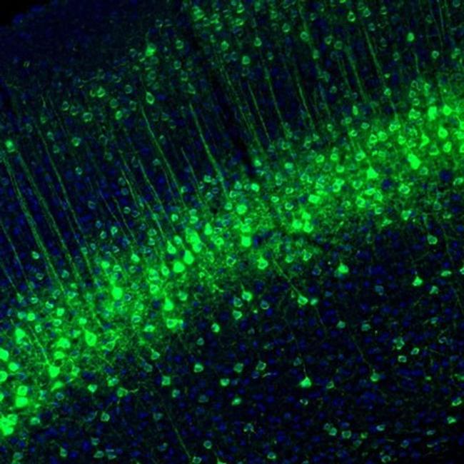
- Immunofluorescent staining of NECAB1 in mouse parietal association cortex shows distinct positivity in layer 4 neurons. Samples were probed using a NECAB1 Polyclonal Antibody (Product # PA5-54849).
- Submitted by
- Invitrogen Antibodies (provider)
- Main image

- Experimental details
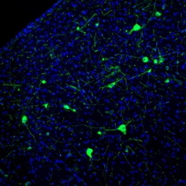
- Immunofluorescent staining of NECAB1 in mouse superior colliculi shows positivity in a subset of neurons. Samples were probed using a NECAB1 Polyclonal Antibody (Product # PA5-54849).

- Submitted by
- Invitrogen Antibodies (provider)
- Main image

- Experimental details
- Immunofluorescent staining of NECAB1 in mouse vestibular nucleus shows immunoreactivity in a subset of neurons. Samples were probed using a NECAB1 Polyclonal Antibody (Product # PA5-54849).
Supportive validation
- Submitted by
- Invitrogen Antibodies (provider)
- Main image

- Experimental details
- Immunohistochemical staining of NECAB1 in human cerebral cortex using NECAB1 Polyclonal Antibody (Product # PA5-54849).

- Submitted by
- Invitrogen Antibodies (provider)
- Main image

- Experimental details
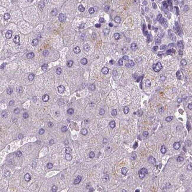
- Immunohistochemical staining of NECAB1 in human liver using NECAB1 Polyclonal Antibody (Product # PA5-54849).
- Submitted by
- Invitrogen Antibodies (provider)
- Main image

- Experimental details
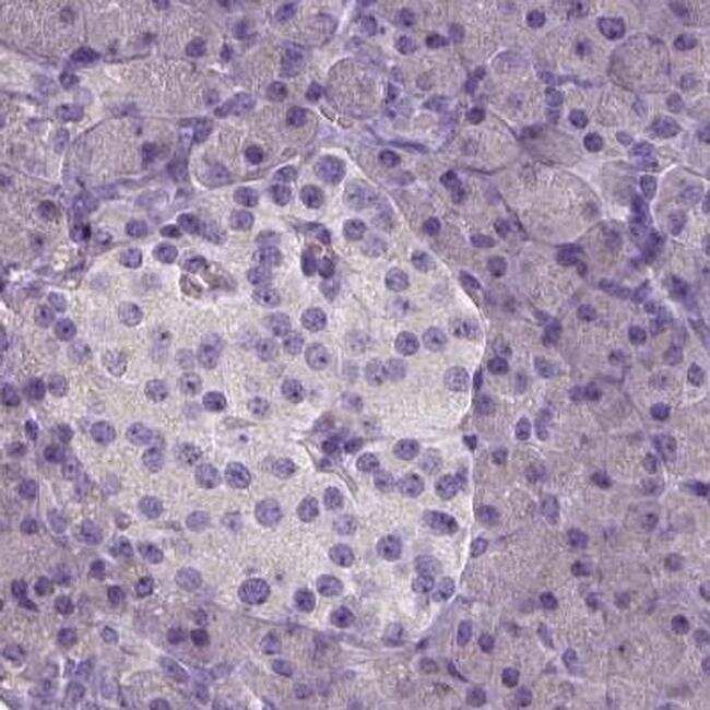
- Immunohistochemical staining of NECAB1 in human pancreas using NECAB1 Polyclonal Antibody (Product # PA5-54849).
- Submitted by
- Invitrogen Antibodies (provider)
- Main image

- Experimental details
- Immunohistochemical staining of NECAB1 in human colon using NECAB1 Polyclonal Antibody (Product # PA5-54849).

- Submitted by
- Invitrogen Antibodies (provider)
- Main image

- Experimental details
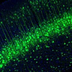
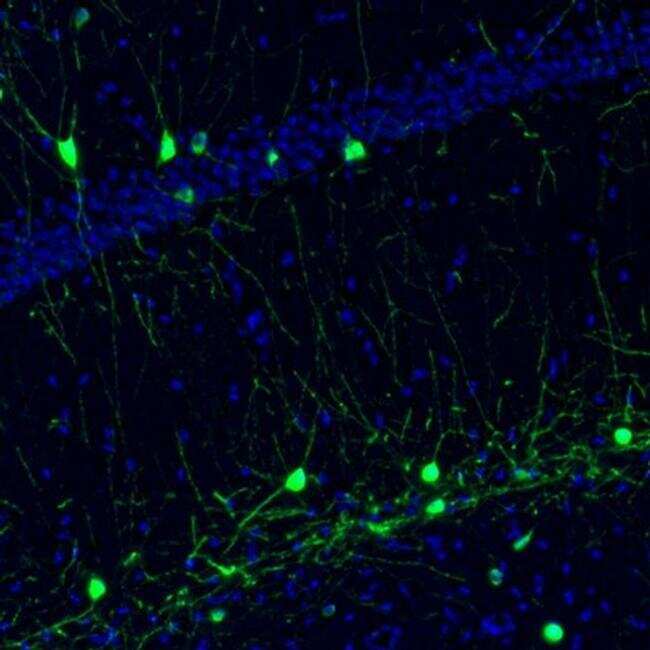
- Immunofluorescent staining of NECAB1 in mouse hippocampus shows immunoreactivity in a subset of interneurons. Samples were probed using a NECAB1 Polyclonal Antibody (Product # PA5-54849).
- Submitted by
- Invitrogen Antibodies (provider)
- Main image

- Experimental details
- Immunohistochemical staining of NECAB1 in human cerebral cortex, colon, liver and pancreas using NECAB1 Polyclonal Antibody (Product # PA5-54849) (A) shows similar protein distribution across tissues to an independent NECAB1 Polyclonal Antibody (B).

Supportive validation
- Submitted by
- Invitrogen Antibodies (provider)
- Main image

- Experimental details
- Fig. 2. Bilateral fibril injection leads to alpha-synuclein inclusion formation in the cortex. Mice received bilateral injections of alpha-syn fibrils at 3-4 months of age. A) pSer129-alpha-synuclein (green; EP1536Y) shows inclusions in coronal sections of the forebrain. Yellow outlines the prefrontal cortex and white outlines other subdivisions of the cortex. B) Double-staining immunofluorescence for NeuN (red) and pSer129-alpha-synuclein (green; EP1536Y) shows banding pattern of inclusions in lower layers (IV/V and VI) of the motor cortex. C) Double-staining immunofluorescence for Necab1 (red) and pSer129-alpha-synuclein (green; 81a) shows localization of inclusions to layer IV/V. Scale bar = 100 mum. Abbreviations: ACC = anterior cingulate cortex, IC = insular cortex, MC = motor cortex, PFC = prefrontal cortex, PLC = prelimbic cortex, SS = somatosensory cortex. (For interpretation of the references to colour in this figure legend, the reader is referred to the web version of this article.)
