Explore
 Validate
 Learn
Learn Western blot
Antibody data
- Antibody Data
- Antigen structure
- References [1]
- Comments [0]
- Validations
- Western blot [3]
- Immunocytochemistry [1]
- Immunohistochemistry [3]
- Other assay [1]
Submit
Validation data
Reference
Comment
Report error
- Product number
- PA5-87443 - Provider product page

- Provider
- Invitrogen Antibodies
- Product name
- PKC delta Polyclonal Antibody
- Antibody type
- Polyclonal
- Antigen
- Recombinant full-length protein
- Reactivity
- Human, Mouse, Rat
- Host
- Rabbit
- Isotype
- IgG
- Vial size
- 100 µL
- Concentration
- 1.12 mg/mL
- Storage
- -20° C, Avoid Freeze/Thaw Cycles
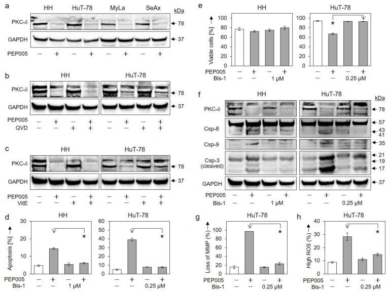
Submitted references Targeting Cutaneous T-Cell Lymphoma Cells by Ingenol Mebutate (PEP005) Correlates with PKCδ Activation, ROS Induction as Well as Downregulation of XIAP and c-FLIP.
Sumarni U, Reidel U, Eberle J
Cells 2021 Apr 23;10(5)
Cells 2021 Apr 23;10(5)
No comments: Submit comment
Supportive validation
- Submitted by
- Invitrogen Antibodies (provider)
- Main image

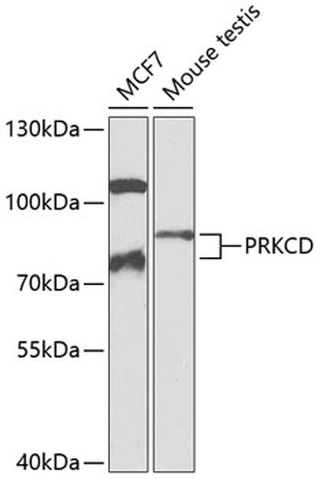
- Experimental details
- Western blot analysis of extracts of various cell lines, using PRKCD Polyclonal antibody (Product # PA5-87443) at 1:1000 dilution. Secondary antibody: HRP Goat Anti-Rabbit IgG (H+L) at 1:10000 dilution. Lysates/proteins: 25ug per lane. Blocking buffer: 3% nonfat dry milk in TBST. Exposure time: 90s.
- Submitted by
- Invitrogen Antibodies (provider)
- Main image
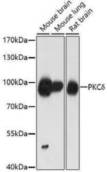
- Experimental details
- Western Blot analysis of PKC delta in extracts of various cell lines using PKC delta Polyclonal Antibody (Product # PA5-87443) at a dilution of 1:1000. A HRP Goat Anti-Rabbit IgG (H+L) secondary antibody was used at a dilution of 1:10,000. Lysates/proteins: 25 µg per lane. Blocking buffer: 3% nonfat dry milk in TBST.
- Submitted by
- Invitrogen Antibodies (provider)
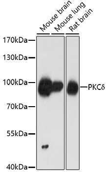
- Main image

- Experimental details
- Western Blot analysis of PKC delta in extracts of various cell lines using PKC delta Polyclonal Antibody (Product # PA5-87443) at a dilution of 1:1000. A HRP Goat Anti-Rabbit IgG (H+L) secondary antibody was used at a dilution of 1:10,000. Lysates/proteins: 25 µg per lane. Blocking buffer: 3% nonfat dry milk in TBST.

Supportive validation
- Submitted by
- Invitrogen Antibodies (provider)
- Main image

- Experimental details
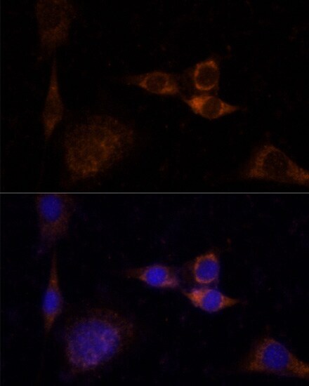
- Immunocytochemistry-Immunofluorescence analysis of PKC delta was performed in NIH/3T3 cells using PKC delta Polyclonal Antibody (Product # PA5-87443).
Supportive validation
- Submitted by
- Invitrogen Antibodies (provider)
- Main image
- Experimental details
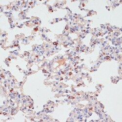
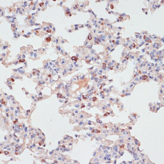
- Immunohistochemistry analysis of PKC delta in paraffin-embedded rat lung using PKC delta Polyclonal Antibody (Product # PA5-87443) at a dilution of 1:100.
- Submitted by
- Invitrogen Antibodies (provider)
- Main image

- Experimental details
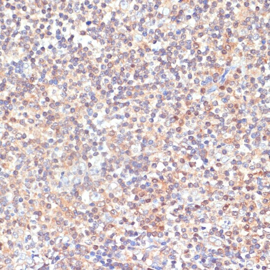
- Immunohistochemistry analysis of PKC delta in paraffin-embedded human tonsil using PKC delta Polyclonal Antibody (Product # PA5-87443) at a dilution of 1:100.
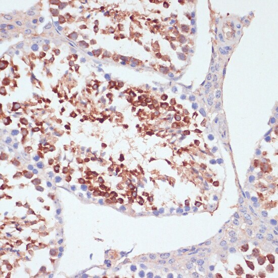
- Submitted by
- Invitrogen Antibodies (provider)
- Main image
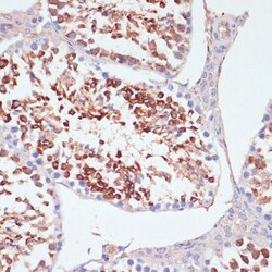
- Experimental details
- Immunohistochemistry analysis of PKC delta in paraffin-embedded mouse testis using PKC delta Polyclonal Antibody (Product # PA5-87443) at a dilution of 1:200.
Supportive validation
- Submitted by
- Invitrogen Antibodies (provider)
- Main image
- Experimental details
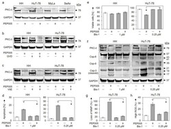
- Figure 6 Role of PKCdelta in PEP005-induced apoptosis. ( a ) Effects of PEP005 (50 nM, 24 h) on PKCdelta proform (78 kDa) were investigated in four CTCL cell lines. ( b , c ) Lacking effects of QVD-Oph (QVD, 5 uM, b) and vitamin E (VitE, 1 mM, c ) on PEP005-induced downregulation of PKCdelta proform are shown (50 nM, 24 h). ( d , e ) Inhibition of PEP005-induced apoptosis ( d ) and restoration of cell viability ( e ) by Bis-1 in HH and HuT-78. Cells were treated for 24 h with PEP005 (50 nM) and/or Bis-1 (HH, 1 uM; HuT-78, 0.25 uM). ( f ) Inhibition of PEP005-mediated caspase-3, -8, and -9 processing through Bis-1, as investigated by Western blotting in HH and HuT-78. Cells were treated for 24 h with 50 nM PEP005; Bis-1 was used at 1 (HH) and 0.25 uM (HuT-78), respectively). ( g , h ) Antagonistic effects of Bis-1 on PEP005-mediated loss of MMP ( g ) and on PEP005-induced ROS production ( h ) in cell line HuT-78 (Time: 24 h; PEP005: 50 nM; Bis-1: 0.25 uM). ( a - c , f ) For Western blotting, 30 ug of each protein extract was loaded per lane, and blots were probed with antibodies for PKCdelta proform (78 kDa), cleaved caspase-3 (21, 19, 17 kDa), caspase-8 (proform, 57 kDa; cleavage products, 43/41 kDa) and caspase-9 (cleavage product, 35 kDa). GAPDH (37 kDa) was used as loading control. For Western blots, two independent series of protein extracts revealed highly comparable results. ( d , e , g , h ) Mean values of triplicates +- SDs of representative experiments are shown. At